. 2010 Oct;38(18):6029-44.
doi: 10.1093/nar/gkq439. Epub 2010 May 27.

Specification of the NF-kappaB transcriptional response by p65 phosphorylation and TNF-induced nuclear translocation of IKK epsilon

Affiliations

Specification of the NF-kappaB transcriptional response by p65 phosphorylation and TNF-induced nuclear translocation of IKK epsilon

Rita Moreno et al. Nucleic Acids Res. 2010 Oct.

Abstract

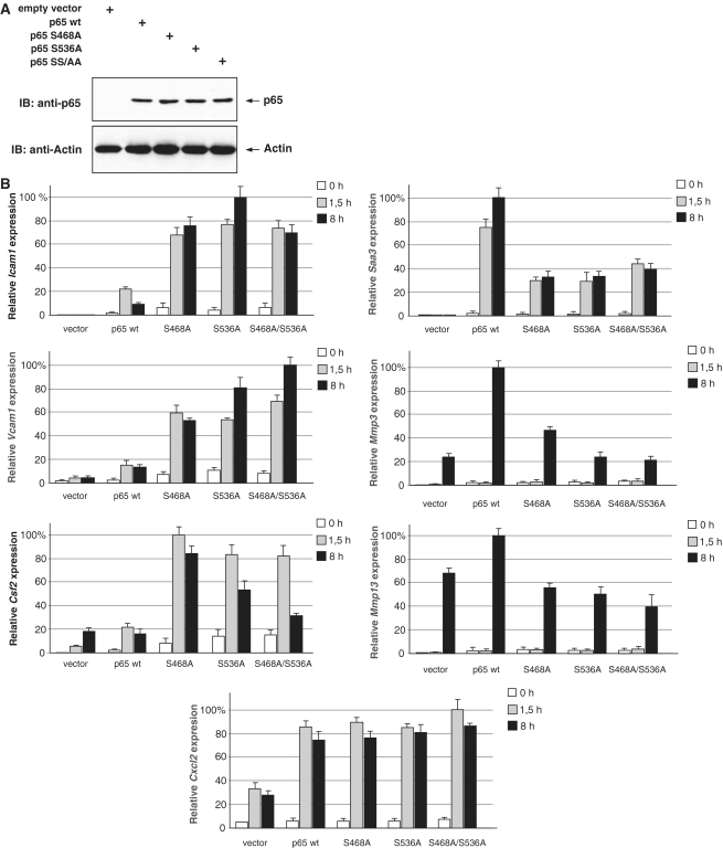
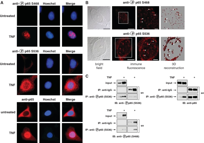
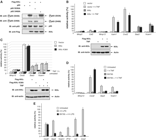
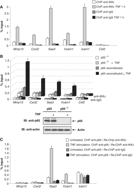
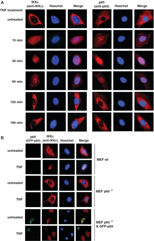
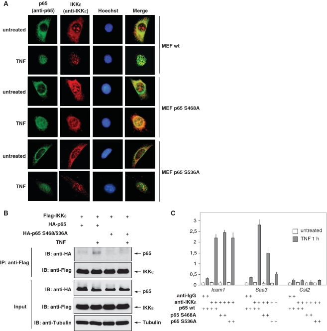
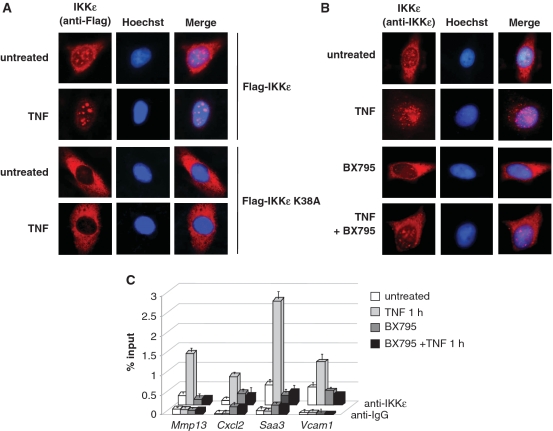
Here we investigated the regulation of NF-κB activity by post-translational modifications upon reconstitution of NF-κB p65-deficient cells with the wild-type protein or phosphorylation-defect mutants. Analysis of NF-κB target gene expression showed that p65 phosphorylations alone or in combination function to direct transcription in a highly target gene-specific fashion, a finding discussed here as the NF-κB barcode hypothesis. High-resolution microscopy and surface rendering revealed serine 536 phosphorylated p65 predominantly in the cytosol, while serine 468 phosphorylated p65 mainly localized in nuclear speckles. TNF stimulation resulted in the translocation of the cytosolic p65 kinase IKKε to the nucleus and also to promyelocytic leukemia (PML) nuclear bodies. This inducible IKKε translocation was dependent on p65 phosphorylation and was prevented by the oncogenic PML-RARα fusion protein. Chromatin immunoprecipitation experiments revealed the inducible association of IKKε to the control regions of several NF-κB target genes. In the nucleus, the kinase contributes to the expression of a subset of NF-κB-regulated genes, thus revealing a novel role of IKKε for the control of nuclear NF-κB activity.


Figures

Figure 1.
Figure 1.
Analysis of phosphorylation-dependent NF-κB p65 activity. (A) p65-deficient MEFs were stably reconstituted to express physiological levels of HA-p65 and its point mutated versions that were changed in serine 468 (p65 S468A), serine 536 (p65 S536A) or both sites (p65 SS/AA). One fraction of the cells was lysed and equal amounts of protein were tested by immunoblotting (IB) for comparable expression of p65. (B) The indicated cells were treated with TNF for 1, 5 or 8 h. The expression of selected NF-κB target genes by real-time PCR. In order to facilitate comparison, maximal gene activation was arbitrarily set as 100%. Experiments were performed in triplicates, error bars display standard deviations.
Figure 2.
Figure 2.
Intracellular distribution of p65 phosphorylation. (A) HeLa cells were left untreated or stimulated with TNF for 30 min. The subcellular localization of endogenous p65 or its phosphorylated forms was visualized by indirect immunofluorescence using anti-phospho-S468, anti-phospho-S536 or anti-p65 antibodies (red). Nuclear DNA was revealed by Hoechst staining. (B) HeLa cells were stimulated for 30 min with TNF and further analyzed by confocal microscopy. A bright field picture, two magnifications of immunofluorescence pictures and a three-dimensional reconstruction of the same cell are displayed. The Z-stack pictures were processed using the Imaris software package for surface rendering. To facilitate the orientation, white arrow heads point to speckles that are displayed by immunofluorescence and also by 3D reconstruction. (C) HeLa cells were stimulated with TNF for 30 min as shown. Lysates were subjected to immunoprecipitation (IP) with anti-IgG control or anti-phospho-p65-S536 antibodies. The input, supernatant (sn) and the immunoprecipitated proteins were further analyzed by western blotting using anti-p65 or phospho-specific antibodies recognizing serine 468 or 536.
Figure 3.
Figure 3.
IKKε-dependent expression of NF-κB target genes. (A) p65-deficient MEFs were transfected with the various expression vectors as shown. Thirty-six hours later they were analyzed by western blotting for expression and phosphorylation of p65 as shown. (B) Ikbke-deficient MEFs were reconstituted to express physiological levels of Flag-IKKε. After 24 h, the cells were treated with TNF for 1 h or left untreated. Gene expression was determined by real-time PCR and normalized to the level of β-actin. Adequate IKKε expression was determined by western blotting using an anti-IKKε antibody. (C) Ikbke−/− MEFs were reconstituted to express Flag-IKKε or the point mutated kinase-inactive version Flag-IKKε-K38A. Cells were further analyzed as in (B). (D) MEFs were treated for 1 h with BX795 (1 μM) or left untreated prior to stimulation with TNF as shown. Gene expression was further quantified by real-time PCR. (E) Primary mouse lung macrophages were preincubated for 1 h with 1 μM BX795 or left untreated prior to stimulation for 4 h with LPS. Gene expression was determined by real-time PCR and normalized to the level of β-actin. All real-time experiments were performed in triplicates, error bars display standard deviations. Maximal gene activation was arbitrarily set as 100%.
Figure 4.
Figure 4.
TNF-induced and NF-κB p65-dependent chromatin recruitment of IKKε. (A) MEFs were stimulated with TNF as shown, followed by ChIP analysis using the indicated specific and anti-IgG control antibodies. IKKε association with different promoter regions was detected by real-time PCR using specific primers. The amounts of p65-associated DNA are presented as the percentage recovered out of the total input DNA (percent input). Experiments were performed in triplicates, error bars display standard deviations. (B) p65-deficient MEFs were transfected with a vector encoding p65 or an adequate control. After 24 h, the cells were stimulated with TNF as shown and ChIP assays for IKKε were performed as in (A). The lower part shows a control western blot ensuring adequate p65 expression. (C) MEFs were stimulated with TNF as shown and used for the ChIP procedure using anti-p65 antibodies or controls, followed by elution and re-ChIP using anti-IKKε antibodies. Experiments were performed in triplicates, error bars display standard deviations.
Figure 5.
Figure 5.
TNF-induced and NF-κB p65-dependent nuclear translocation of IKKε. (A) HeLa cells were stimulated with TNF for the indicated periods. The intracellular localization of endogenous IKKε and p65 proteins was analyzed by indirect immunofluorescence using anti-p65 and anti-IKKε antibodies (red). Nuclear DNA was revealed by Hoechst staining. (B) Wild-type MEFs and p65-deficient MEFs transfected with empty vector or a plasmid encoding GFP-p65 were stimulated for 30 min with TNF as shown. The subcellular localization of GFP-p65 (green) and of endogenous IKKε (red) was analyzed by indirect immunofluorescence.
Figure 6.
Figure 6.
Phosphorylation-dependent association of IKKε and p65. (A) MEFs stably reconstituted with p65 wild-type or the point mutated versions p65 S468A and p65 S536A were stimulated with TNF and the subcellular localization of IKKε (red) and p65 (green) was visualized by indirect immunofluorescence. (B) HEK293 cells transfected to express Flag-IKKε together with HA-p65 or its point mutated version HA-p65 SS/AA were stimulated with TNF, lysed and cell extracts subjected to immunoprecipitation as shown. The input material and the immunoprecipitated proteins were further analyzed by western blotting using anti-HA and anti-Flag antibodies. (C) MEFs stably reconstituted with p65 wild-type or the point mutated versions p65 S468A and p65 S536A were stimulated with TNF for 1 h, followed by ChIP analysis using the indicated specific and anti-IgG control antibodies. IKKε association with the indicated promoter regions was detected by real-time PCR using specific primers, error bars show standard deviations from three experiments.
Figure 7.
Figure 7.
Nuclear import and chromatin association of IKKε depends on its kinase activity. (A) Ikbke-deficient MEFs were reconstituted to express Flag-IKKε or Flag-IKKε-K38A. Thirty-six hours after transfection, cells were left untreated or stimulated with TNF for 30 min. The subcellular localization of IKKε was visualized by indirect immunofluorescence using an anti-Flag antibody (red). (B) MEFs were incubated for 1 h with BX795 (1 μM) as shown and then further treated for 30 min with TNF. Subcellular localization of endogenous IKKε was visualized by indirect immunofluorescence using an anti-IKKε antibody. (C) MEFs were preincubated for 1 h with 1 μM BX795 or left untreated prior to stimulation for 1 h with TNF. ChIP assays for IKKε were performed using specific anti-IKKε and anti-IgG control antibodies and the precipitated DNA was quantified by real-time PCR using specific primers. Experiments were performed in triplicates, error bars display standard deviations.
Figure 8.
Figure 8.
TNF-induced recruitment of IKKε to PML-NBs. (A) HeLa cells were transfected to express the PML splicing forms PML-III, PML-IV or the PML-RARα fusion protein. Thirty-six hours after the transfection, cells were left untreated or stimulated with TNF for 30 min. The subcellular localization of PML and endogenous IKKε was visualized by indirect immunofluorescence using an anti-PML (red) and anti-IKKε antibody (green). (B) Upper: Hela cells transfected with a plasmid expressing a PML-specific shRNA were treated with TNF for 30 min as shown and analyzed for subcellular localization of endogenous IKKε using an anti-IKKε antibody. Lower: the efficient knock-down of PML was controlled by transfection of the vector directing the synthesis of the PML specific shRNA and detection of PML by immunofluorescence. The limited transfection efficiency allows to observe PML down-regulation only in transfected cells. (C) Cells were transfected with a vector for a PML specific shRNA or a control. After 36 h, cells were stimulated for 30 min with TNF as shown and the localization of the endogenous proteins was revealed by immunofluorescence. (D) Hela cells were treated as in (C) and analyzed for the occurrence and subcellular localization of endogenous PML and serine 468 phosphorylated p65 as shown. (E) Schematic model summarizing the mechanisms and functions of p65 phosphorylation and its phosphorylation by IKKε and other kinases.

Similar articles

Cited by

References

    1. Gaestel M, Kotlyarov A, Kracht M. Targeting innate immunity protein kinase signalling in inflammation. Nat. Rev. Drug Discov. 2009;8:480–499. - PubMed
    1. Vallabhapurapu S, Karin M. Regulation and function of NF-kappaB transcription factors in the immune system. Annu. Rev. Immunol. 2009;27:693–733. - PubMed
    1. Natoli G, De Santa F. Shaping alternative NF-kappaB-dependent gene expression programs: new clues to specificity. Cell Death Differ. 2006;13:693–696. - PubMed
    1. Hayden MS, Ghosh S. Shared principles in NF-kappaB signaling. Cell. 2008;132:344–362. - PubMed
    1. Hacker H, Karin M. Regulation and function of IKK and IKK-related kinases. Sci. STKE. 2006;2006:re13. - PubMed

Publication types