. 2010 Jul 15;16(14):3639-47.
doi: 10.1158/1078-0432.CCR-09-3385. Epub 2010 May 27.

Augmentation of radiation response by motesanib, a multikinase inhibitor that targets vascular endothelial growth factor receptors

Affiliations

Augmentation of radiation response by motesanib, a multikinase inhibitor that targets vascular endothelial growth factor receptors

Tim J Kruser et al. Clin Cancer Res. .

Abstract

Background: Motesanib is a potent inhibitor of vascular endothelial growth factor receptors (VEGFR) 1, 2, and 3, platelet-derived growth factor receptor, and Kit receptors. In this report we examine the interaction between motesanib and radiation in vitro and in head and neck squamous cell carcinoma (HNSCC) xenograft models.

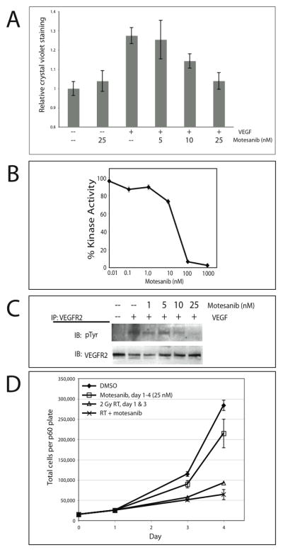
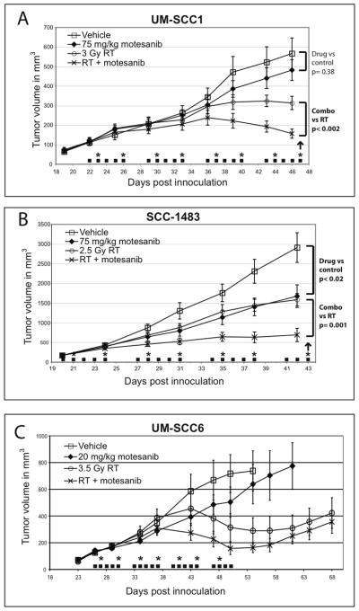
Experimental design: In vitro assays were done to assess the impact of motesanib on VEGFR2 signaling pathways in human umbilical vein endothelial cells (HUVEC). HNSCC lines grown as tumor xenografts in athymic nude mice were utilized to assess the in vivo activity of motesanib alone and in combination with radiation.

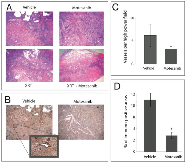
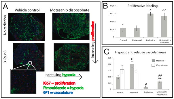
Results: Motesanib inhibited VEGF-stimulated HUVEC proliferation in vitro, as well as VEGFR2 kinase activity. Additionally, motesanib and fractionated radiation showed additive inhibitory effects on HUVEC proliferation. In vivo combination therapy with motesanib and radiation showed increased response compared with drug or radiation alone in UM-SCC1 (P < 0.002) and SCC-1483 xenografts (P = 0.001); however, the combination was not significantly more efficacious than radiation alone in UM-SCC6 xenografts. Xenografts treated with motesanib showed a reduction of vessel penetration into tumor parenchyma, compared with control tumors. Furthermore, triple immunohistochemical staining for vasculature, proliferation, and hypoxia showed well-defined spatial relationships among these parameters in HNSCC xenografts. Motesanib significantly enhanced intratumoral hypoxia in the presence and absence of fractionated radiation.

Conclusions: These studies identify a favorable interaction when combining radiation and motesanib in HNSCC models. The data presented suggest that motesanib reduces blood vessel penetration into tumors and thereby increases intratumoral hypoxia. These findings suggest that clinical investigations examining combinations of radiation and motesanib are warranted in HNSCC.


Conflict of interest statement

Conflict of Interest Notification: AC and AP are employees of Amgen, Inc.

Figures

Fig 1
Fig 1
Motesanib in vitro activity on VEGFR2 signaling and interaction with radiation. (A) Impact of motesanib on VEGF-stimulated HUVEC proliferation. Cells were grown in EBM-2 basal medium with 2% FBS, without exogenous growth factors. VEGF stimulation was 25 ng/ml every 24 hours x 4 days. Final DMSO concentration was 0.25% in all wells. Data points represent the mean crystal violet staining intensity (6 wells per condition) +/− SEM. (B) Motesanib blocks VEGFR2 kinase activity. VEGFR2 kinase activity was determined in presence of serial dilutions of motesanib. (C) Motesanib blocks VEGF stimulation of VEGFR2 phosphorylation in HUVECs. HUVECs were pretreated with motesanib for 24 hours, collected after stimulation with 50 ng/ml VEGF x 45 min. IP = immunoprecipitation; IB = immunoblot. (D) HUVECs seeded day 0, exposed to motesanib or DMSO days 1–4, and radiated x 2 on days 1 and 3 were harvested and counted via trypan blue exclusion. Points represent mean of 3 plates per condition at days 1, 3, and 4, +/− SEM
Fig 2
Fig 2
Motesanib augments radiation response in tumor xenograft models. Mice bearing UM-SCC1 (A), SCC-1483 (B), or UMSCC-6 (C) tumors were treated with either motesanib or vehicle by oral gavage 5x weekly (■). Twice weekly radiation was also administered (*). Data points are expressed as mean tumor size (n=10/group) +/− SEM. Arrows represent days that tumors were harvested for immunohistochemistry (as presented in Figures 3 & 4).
Fig 3
Fig 3
Vascular distribution and tumor architecture in UM-SCC1 xenografts. Tumors from mice harboring UM-SCC1 xenografts were harvested after 4 weeks of treatment (see Fig. 2 for details). Tumor architecture (A) was observed under low power (20x) via H&E staining. (B) Tumor tissue was stained for expression of von Willebrand Factor (vWF), which stains tumor vasculature, as demonstrated at 200x (inset). (C,D) Quantitative analysis of vWF staining was performed at 100x magnification. Five high-powered fields per tumor were randomly chosen and vessel density was quantified by counting the vessels (C) and examining the percentage of vWF immunoreactive area (D) using Image J software. Control tumors had a significantly larger proportion staining positive for vWF than motesanib-treated tumors (* p < 0.01; Students t-test). Data represent mean ± SEM.
Fig 4
Fig 4
Impact of motesanib on intratumoral hypoxia (pimonidazole), proliferation (Ki67), and vasculature (9F1). Mice bearing UMSCC-1 xenografts (see Fig. 2 for treatment schema) were inoculated with pimonidazole (PIMO) by intra peritoneal injection 1 hour after the last radiation dose. Tumors were then harvested 2 hours later (3 hours after last radiation dose). Tumor tissue was then analyzed by immunohistochemistry for expression of PIMO, 9F1, and Ki67. Representative tumor sections are shown. (A) Tumor hypoxia and proliferation show spatial relationships to tumor vasculature (inset) (B) Four weeks of fractionated radiation results in higher levels of proliferative staining (Ki67), both in the absence of motesanib (^; p=0.02) and in the presence of motesanib (^^; p=0.04) (Fig 4B). Motesanib was not demonstrated to impact tumor cell proliferation. (C) Hypoxic staining of each tumor was analyzed, and impact of motesanib on vascular staining was assessed. Treatment with motesanib resulted in significantly more hypoxia in tumors not exposed to fractionated radiation (*; p <0.02, students t-test) and tumors exposed to fractionated radiation (**; p <0.01). Radiation significantly reduced the hypoxic fraction in tumors in the absence (#; p <0.01) and presence (##; p <0.01) of motesanib. The impact of motesanib on tumor vasculature was not apparent in this subset of xenografts Data represent mean ± SEM (n=3 in all groups).

Similar articles

Cited by

References

    1. Jemal A, Siegel R, Ward E, et al. Cancer statistics, 2008. CA Cancer J Clin. 2008;58:71–96. - PubMed
    1. Pignon JP, le Maitre A, Bourhis J. Meta-Analyses of Chemotherapy in Head and Neck Cancer (MACH-NC): an update. Int J Radiat Oncol Biol Phys. 2007;69:S112–4. - PubMed
    1. Bonner JA, Harari PM, Giralt J, et al. Radiotherapy plus cetuximab for squamous-cell carcinoma of the head and neck. N Engl J Med. 2006;354:567–78. - PubMed
    1. Kyzas PA, Stefanou D, Batistatou A, Agnantis NJ. Prognostic significance of VEGF immunohistochemical expression and tumor angiogenesis in head and neck squamous cell carcinoma. J Cancer Res Clin Oncol. 2005;131:624–30. - PubMed
    1. Oc P, Rhys-Evans P, Eccles SA. Expression of vascular endothelial growth factor family members in head and neck squamous cell carcinoma correlates with lymph node metastasis. Cancer. 2001;92:556–68. - PubMed

Publication types

MeSH terms