. 2010 Aug 6;285(32):24519-28.
doi: 10.1074/jbc.M110.125450. Epub 2010 May 27.

Crystal structure of exotype alginate lyase Atu3025 from Agrobacterium tumefaciens

Affiliations

Crystal structure of exotype alginate lyase Atu3025 from Agrobacterium tumefaciens

Akihito Ochiai et al. J Biol Chem. .

Abstract

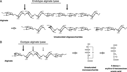
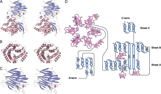
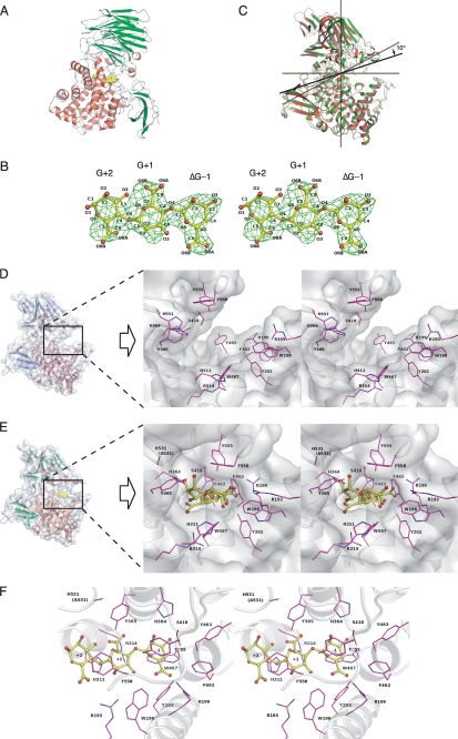
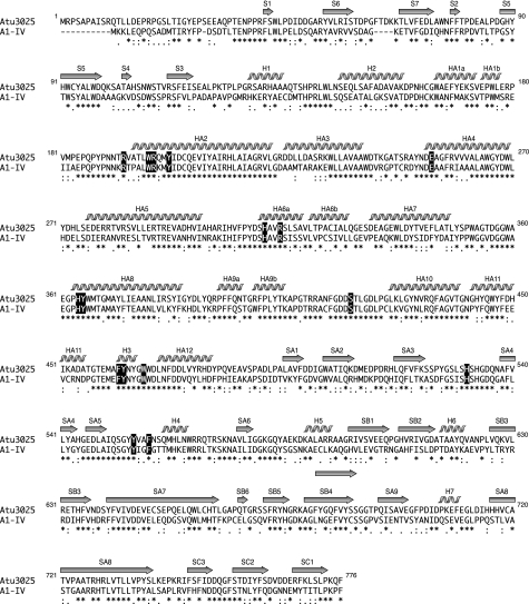
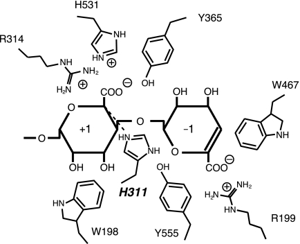
Alginate, a major component of the cell wall matrix in brown seaweeds, is degraded by alginate lyases through a beta-elimination reaction. Almost all alginate lyases act endolytically on substrate, thereby yielding unsaturated oligouronic acids having 4-deoxy-l-erythro-hex-4-enepyranosyluronic acid at the nonreducing end. In contrast, Agrobacterium tumefaciens alginate lyase Atu3025, a member of polysaccharide lyase family 15, acts on alginate polysaccharides and oligosaccharides exolytically and releases unsaturated monosaccharides from the substrate terminal. The crystal structures of Atu3025 and its inactive mutant in complex with alginate trisaccharide (H531A/DeltaGGG) were determined at 2.10- and 2.99-A resolutions with final R-factors of 18.3 and 19.9%, respectively, by x-ray crystallography. The enzyme is comprised of an alpha/alpha-barrel + anti-parallel beta-sheet as a basic scaffold, and its structural fold has not been seen in alginate lyases analyzed thus far. The structural analysis of H531A/DeltaGGG and subsequent site-directed mutagenesis studies proposed the enzyme reaction mechanism, with His(311) and Tyr(365) as the catalytic base and acid, respectively. Two structural determinants, i.e. a short alpha-helix in the central alpha/alpha-barrel domain and a conformational change at the interface between the central and C-terminal domains, are essential for the exolytic mode of action. This is, to our knowledge, the first report on the structure of the family 15 enzyme.


Figures

FIGURE 1.
FIGURE 1.
Alginate degradation by endotype (A) and exotype (B) alginate lyases. Thick arrows indicate the cleavage sites for each enzyme against substrate.
FIGURE 2.
FIGURE 2.
Structure of Atu3025. A, overall structure (stereodiagram). B, central α-domain (stereodiagram). C, C-terminal β-domain (stereodiagram). D, topology diagram. β-Sheets are shown as blue arrows, and helices are shown as pink cylinders.
FIGURE 3.
FIGURE 3.
Active site structure of Atu3025. A, overall structure of H531A/ΔGGG. B, electron density of the ΔGGG molecule in the active pocket by the omit map (FoFc) calculated without ΔGGG and countered at 3.0 σ. C, superimposition of ligand-free Atu3025 (red) and H531A/ΔGGG (green). D, surface model and active site structure (inset, stereodiagram) of the ligand-free Atu3025. E, surface model and active site structure (inset, stereodiagram) of H531A/ΔGGG. F, residues in the H531A/ΔGGG structure interacting with the ΔGGG molecule in the active pocket. Amino acid residues and ΔGGG molecule are shown by colored elements: oxygen atom, red, carbon atom, pink in amino acid residues, and yellow in the ΔGGG molecule; nitrogen atom, deep blue. Characters in panel B indicate the saccharide number and its atoms. Characters in panels D–F indicate the subsite and amino acid residue numbers.
FIGURE 4.
FIGURE 4.
Alignment of amino acid sequences of family PL-15 alginate lyase. Atu3025, alginate lyase from A. tumefaciens strain C58 (GenPept accession number AAL43841); A1-IV, alginate lyase from Sphingomonas sp. strain A1 (GenPept accession number BAB03319). Amino acid sequences were aligned using the ClustalW program. Identical and similar amino acid residues in these alginate lyases are denoted by asterisks and dots, respectively. Secondary structure elements of Atu3025 are as shown above. Amino acid residues interacting with ΔGGG are highlighted in black.
FIGURE 5.
FIGURE 5.
Catalytic center in Atu3025. The residues surrounding subsites −1 and +1 are represented schematically.

References

    1. Cantarel B. L., Coutinho P. M., Rancurel C., Bernard T., Lombard V., Henrissat B. (2009) Nucleic Acids Res. 37, D233–D238 - PMC - PubMed
    1. Wong T. Y., Preston L. A., Schiller N. L. (2000) Annu. Rev. Microbiol. 54, 289–340 - PubMed
    1. Yoon H.-J., Hashimoto W., Miyake O., Okamoto M., Mikami B., Murata K. (2000) Protein Expr. Purif. 19, 84–90 - PubMed
    1. Hashimoto W., Miyake O., Momma K., Kawai S., Murata K. (2000) J. Bacteriol. 182, 4572–4577 - PMC - PubMed
    1. Ochiai A., Hashimoto W., Murata K. (2006) Res. Microbiol. 157, 642–649 - PubMed

Publication types

LinkOut - more resources