. 2010 Jul 30;285(31):23647-54.
doi: 10.1074/jbc.M110.127050. Epub 2010 May 27.

The activity of hyaluronan synthase 2 is regulated by dimerization and ubiquitination

Affiliations

The activity of hyaluronan synthase 2 is regulated by dimerization and ubiquitination

Eugenia Karousou et al. J Biol Chem. .

Abstract

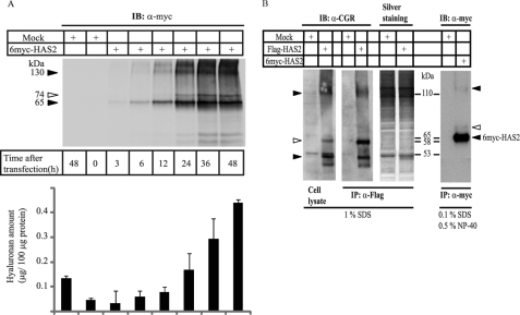
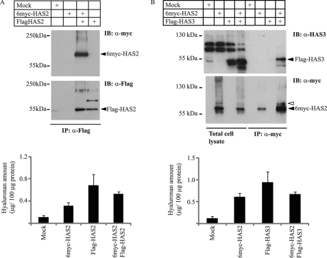
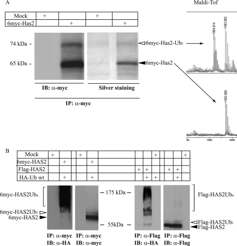
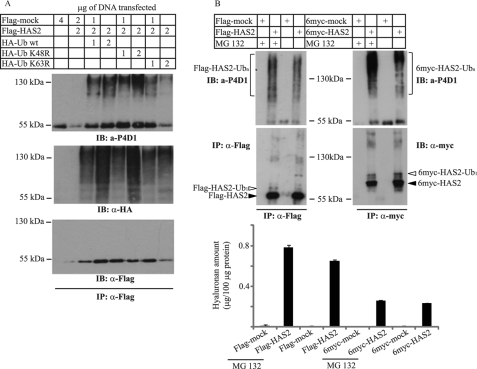
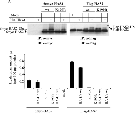
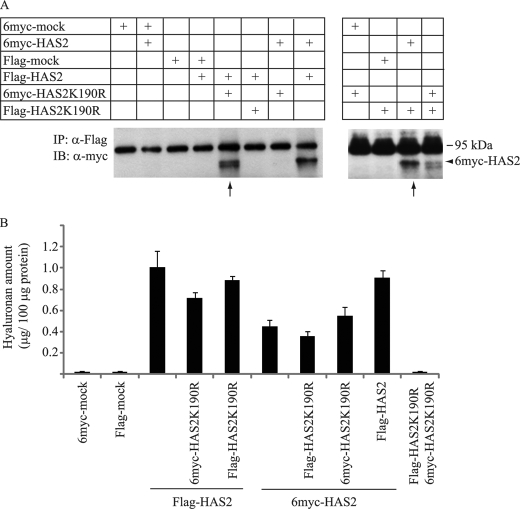
Hyaluronan is a component of the extracellular matrix, which affects tissue homeostasis. In this study, we investigated the regulatory mechanisms of one of the hyaluronan-synthesizing enzymes, HAS2. Ectopic expression of Flag- and 6myc-HAS2 in COS-1 cells followed by immunoprecipitation and immunoblotting revealed homodimers; after co-transfection with Flag-HAS3, also heterodimers were seen. Furthermore, the expressed HAS2 was ubiquitinated. We identified one acceptor site for ubiquitin on lysine residue 190. Mutation of this residue led to inactivation of the enzymatic activity of HAS2. Interestingly, K190R-mutated HAS2 formed dimers with wt HAS2 and quenched the activity of wt HAS2, thus demonstrating a functional role of the dimeric configuration.


Figures

FIGURE 1.
FIGURE 1.
Structure and activity of HAS2 expressed in COS-1 cells. A, membrane fractions from COS-1 cells transiently transfected with 6myc-HAS2 or with empty vector (mock), were solubilized in TBS/Ca2+ buffer supplemented with 1% SDS. Total cell lysates were separated by SDS-PAGE followed by immunoblotting (IB) with Myc antibodies to detect the expression of 6myc-HAS2. The hyaluronan content in conditioned media at different time points was determined as described under “Materials and Methods.” The error bars indicate ranges of duplicate samples. B, membrane fractions from COS-1 cells transiently transfected with 6myc-HAS2, Flag-HAS2, or transfected with empty vectors were solubilized in TBS/Ca2+ buffer supplemented with 1% SDS or 0.1% SDS and 0.5% Nonidet P-40. Total cell lysates or immunoprecipitated (IP) material were separated by SDS-PAGE, and the gels were subjected to silver-staining or IB, as described under “Materials and Methods.” Immunoreactive bands were visualized with antibodies against HAS2 (α-CGR) and myc. Filled arrowheads indicate the position of oligomeric- and polymeric-tagged HAS2 species. Open arrowheads indicate a slower migrating species of tagged HAS2. Data in panels A and B represent one of two and one of three experiments with similar results, respectively.
FIGURE 2.
FIGURE 2.
6myc-HAS2 forms homo-oligomers and hetero-oligomers with Flag-HAS3. 6myc-HAS2, Flag-HAS2, and/or Flag-HAS3 were transiently transfected into COS-1 cells. Lysates were subjected to immunoprecipitation with antibodies against Flag- or 6myc-HAS2, samples were resolved by SDS-PAGE and possible homo-oligomerization (A) as well as hetero-oligomerization (B) were analyzed with immunoblotting with anti-Myc or anti-Flag antibodies, as well as with an antiserum against HAS3. The hyaluronan content in the 48 h conditioned media was measured as described under “Materials and Methods.” Data in panels A and B are the mean of duplicate ± variation from a representative experiment out of two with similar results.
FIGURE 3.
FIGURE 3.
Mono- and polyubiquitination of HAS2 expressed by COS-1 cells. COS-1 cells were transfected with 6myc-HAS2, Flag-HAS2, or a mock construct in the absence or presence of HA-Ub. A, lysates were subjected to immunoprecipitation by Myc antibody followed by SDS-PAGE and immunoblot analysis with antibodies against Myc or silver staining. The immunoreactive bands of 65 and 74 kDa were cut out from the silver-stained gel and analyzed with MALDI-TOF. B, immunoprecipitation was also performed with antibodies against myc-HAS2 (myc) or Flag-HAS2 (Flag), followed by immunoblot analysis with Myc or Flag as well as with antibodies against HA-Ub (HA). Data in A and B are representative experiments out of two and three experiments with similar results, respectively.
FIGURE 4.
FIGURE 4.
Effects of MG132 on polyubiquitination and activity of tagged HAS2. A, Flag-pcDNA3 vector and Flag-HAS2 alone or together with HA-Ub (either wt or the K48R and K63R Ub mutants) were expressed in COS-1 cells at different amounts. Cell extracts were subjected to immunoprecipitation with antibodies against Flag-HAS2 (Flag) and sequential immunoblot analysis with antibodies against HA-Ub (P4D1), Flag-HAS2 (Flag), and anti-HA. B, mock-transfected and Flag- or 6myc-HAS2 transfected cells were treated with the proteasomal inhibitor MG132 (25 μm) for 8 h. Cell lysates were subjected to immunoprecipitation with antibodies against Flag-HAS2 (Flag) and 6myc-HAS2 (myc), followed by immunoblot analysis with antibodies against tagged HAS2, then the membranes were stripped and reprobed with anti-HA-Ub (P4D1). Conditioned media, after 48 h of incubation, were analyzed for hyaluronan content as described under “Materials and Methods.” Each value depicts mean of triplicates ± S.D. Data in A and B represent one of three experiments performed with similar results.
FIGURE 5.
FIGURE 5.
Lys-190 in Flag- and 6myc-HAS2 is monoubiquitinated and crucial for HAS2 enzymatic activity. A, COS-1 cells were transfected with 2 μg of either 6myc- or Flag-HAS2 constructs alone, as well as with 2 μg of their respective K190R mutants. Some cell cultures were also transfected with wt HA-Ub (1 μg). Samples were subjected to immunoprecipitation with antibodies against 6myc-HAS2 (myc) or Flag-HAS2 (Flag), followed by immunoblot analysis with antibodies against Myc and Flag, respectively. Filled arrowheads indicate the position of ubiquitinated-tagged HAS2 species. Open arrowheads point out the slower migrating monoubiquitinated forms (Ub1). B, conditioned media, after 48 h of transfection, were analyzed for hyaluronan content, as described under “Materials and Methods.” Each value depicts means of duplicates ± variation. Data in A and B represent one of three experiments performed with similar results.
FIGURE 6.
FIGURE 6.
HAS2 K190R mutants can form oligomers with wt HAS2. A, COS-1 cells were transfected with 2 μg of either wt 6myc- or Flag-HAS2 constructs with or without 2 μg of their respective K190R mutants. Samples were then subjected to immunoprecipitation with antibodies against Flag-HAS2 (Flag), followed by immunoblot analysis with antibodies against Myc. Thin arrows indicate co-immunoprecipitation of wt 6myc- and Flag-HAS2 with K190R mutants. B, bar graph shows the hyaluronan content in 48 h conditioned media of cells transfected with the indicated vectors. Each value depicts the means of triplicates ± S.D. Data in A and B, are representative experiments out of four experiments performed with similar results.
FIGURE 7.
FIGURE 7.
Increased expression of Flag-HAS2 K190R suppresses the HA-HAS2-induced hyaluronan production. COS-1 cells were transfected with 0.5 μg of HA-wt HAS2, Flag-HAS2 K190R, HA-Ub, or mock alone. Some of the HA-HAS2 transfected cultures were co-transfected with increasing amounts of Flag-HAS2 K190R constructs (0.5–6 μg) without or together with 0.5 μg of wt HA-Ub. Conditioned media, after 48 h of transfection, were analyzed for hyaluronan content as described under “Materials and Methods.” Each value depicts means of triplicates ± S.D. The data shown are representative of one out of four experiments with similar results.

Similar articles

Cited by

References

    1. Laurent T. C., Fraser J. R. E. (1992) FASEB J. 6, 2397–2404 - PubMed
    1. Stuhlmeier K. M. (2006) Wien. Med. Wochenschr. 156, 563–568 - PubMed
    1. Toole B. P. (2004) Nat. Rev. Cancer 4, 528–539 - PubMed
    1. Toole B. P., Slomiany M. G. (2008) Semin. Cancer Biol. 18, 244–250 - PMC - PubMed
    1. Itano N., Kimata K. (2008) Semin. Cancer Biol. 18, 268–274 - PubMed

Publication types