

Site mapping and characterization of O-glycan structures on alpha-dystroglycan isolated from rabbit skeletal muscle
- PMID: 20507986
- PMCID: PMC2915724
- DOI: 10.1074/jbc.M110.126474
Site mapping and characterization of O-glycan structures on alpha-dystroglycan isolated from rabbit skeletal muscle
Abstract
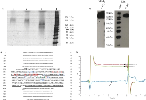
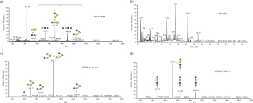
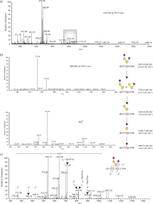
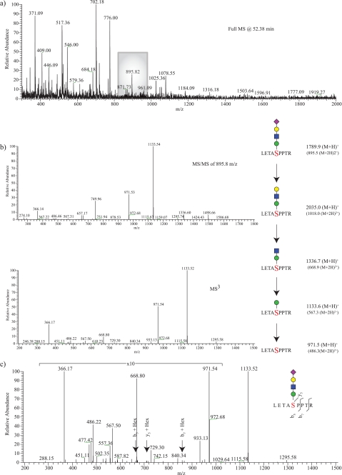
The main extracellular matrix binding component of the dystrophin-glycoprotein complex, alpha-dystroglycan (alpha-DG), which was originally isolated from rabbit skeletal muscle, is an extensively O-glycosylated protein. Previous studies have shown alpha-DG to be modified by both O-GalNAc- and O-mannose-initiated glycan structures. O-Mannosylation, which accounts for up to 30% of the reported O-linked structures in certain tissues, has been rarely observed on mammalian proteins. Mutations in multiple genes encoding defined or putative glycosyltransferases involved in O-mannosylation are causal for various forms of congenital muscular dystrophy. Here, we explore the glycosylation of purified rabbit skeletal muscle alpha-DG in detail. Using tandem mass spectrometry approaches, we identify 4 O-mannose-initiated and 17 O-GalNAc-initiated structures on alpha-DG isolated from rabbit skeletal muscle. Additionally, we demonstrate the use of tandem mass spectrometry-based workflows to directly analyze glycopeptides generated from the purified protein. By combining glycomics and tandem mass spectrometry analysis of 91 glycopeptides from alpha-DG, we were able to assign 21 different residues as being modified by O-glycosylation with differing degrees of microheterogeneity; 9 sites of O-mannosylation and 14 sites of O-GalNAcylation were observed with only two sites definitively exhibiting occupancy by either type of glycan. The distribution of identified sites of O-mannosylation suggests a limited role for local primary sequence in dictating sites of attachment.
Figures
Similar articles
-
Glycomic analyses of mouse models of congenital muscular dystrophy.J Biol Chem. 2011 Jun 17;286(24):21180-90. doi: 10.1074/jbc.M110.203281. Epub 2011 Apr 1. J Biol Chem. 2011. PMID: 21460210 Free PMC article.
-
Characterization of site-specific O-glycan structures within the mucin-like domain of alpha-dystroglycan from human skeletal muscle.Glycobiology. 2010 Sep;20(9):1160-9. doi: 10.1093/glycob/cwq082. Epub 2010 May 27. Glycobiology. 2010. PMID: 20507882
-
Carbohydrate-binding domain of the POMGnT1 stem region modulates O-mannosylation sites of α-dystroglycan.Proc Natl Acad Sci U S A. 2016 Aug 16;113(33):9280-5. doi: 10.1073/pnas.1525545113. Epub 2016 Aug 4. Proc Natl Acad Sci U S A. 2016. PMID: 27493216 Free PMC article.
-
O-Mannosylation and human disease.Cell Mol Life Sci. 2013 Aug;70(16):2849-57. doi: 10.1007/s00018-012-1193-0. Epub 2012 Nov 1. Cell Mol Life Sci. 2013. PMID: 23115008 Free PMC article. Review.
-
Dissecting the molecular basis of the role of the O-mannosylation pathway in disease: α-dystroglycan and forms of muscular dystrophy.Chembiochem. 2013 Dec 16;14(18):2392-402. doi: 10.1002/cbic.201300417. Epub 2013 Nov 7. Chembiochem. 2013. PMID: 24318691 Free PMC article. Review.
Cited by
-
Glycomic analyses of mouse models of congenital muscular dystrophy.J Biol Chem. 2011 Jun 17;286(24):21180-90. doi: 10.1074/jbc.M110.203281. Epub 2011 Apr 1. J Biol Chem. 2011. PMID: 21460210 Free PMC article.
-
B4GAT1 is the priming enzyme for the LARGE-dependent functional glycosylation of α-dystroglycan.Elife. 2014 Oct 3;3:e03943. doi: 10.7554/eLife.03943. Elife. 2014. PMID: 25279697 Free PMC article.
-
Transgenic overexpression of LARGE induces α-dystroglycan hyperglycosylation in skeletal and cardiac muscle.PLoS One. 2010 Dec 28;5(12):e14434. doi: 10.1371/journal.pone.0014434. PLoS One. 2010. PMID: 21203384 Free PMC article.
-
Glycoproteomic characterization of recombinant mouse α-dystroglycan.Glycobiology. 2012 May;22(5):662-75. doi: 10.1093/glycob/cws002. Epub 2012 Jan 11. Glycobiology. 2012. PMID: 22241827 Free PMC article.
-
Structural glycomic analyses at high sensitivity: a decade of progress.Annu Rev Anal Chem (Palo Alto Calif). 2013;6:237-65. doi: 10.1146/annurev-anchem-062012-092609. Epub 2013 Apr 3. Annu Rev Anal Chem (Palo Alto Calif). 2013. PMID: 23560930 Free PMC article. Review.
References
-
- Jaeken J., Hennet T., Freeze H. H., Matthijs G. (2008) J. Inherited Metab. Dis. 31, 669–672 - PubMed
-
- Aebi M., Helenius A., Schenk B., Barone R., Fiumara A., Berger E. G., Hennet T., Imbach T., Stutz A., Bjursell C., Uller A., Wahlström J. G., Briones P., Cardo E., Clayton P., Winchester B., Cormier-Dalre V., de Lonlay P., Cuer M., Dupré T., Seta N., de Koning T., Dorland L., de Loos F., Kupers L. (1999) Glycoconj. J. 16, 669–671 - PubMed
-
- Cohn R. D. (2005) Neuromuscul. Disord. 15, 207–217 - PubMed
-
- Haltiwanger R. S., Lowe J. B. (2004) Annu. Rev. Biochem. 73, 491–537 - PubMed
-
- Barresi R., Campbell K. P. (2006) J. Cell Sci. 119, 199–207 - PubMed
Publication types
MeSH terms
Substances
Grants and funding
LinkOut - more resources
Full Text Sources
Other Literature Sources
