. 2010 Aug 6;285(32):24845-54.
doi: 10.1074/jbc.M110.128546. Epub 2010 May 27.

Microvirin, a novel alpha(1,2)-mannose-specific lectin isolated from Microcystis aeruginosa, has anti-HIV-1 activity comparable with that of cyanovirin-N but a much higher safety profile

Affiliations

Microvirin, a novel alpha(1,2)-mannose-specific lectin isolated from Microcystis aeruginosa, has anti-HIV-1 activity comparable with that of cyanovirin-N but a much higher safety profile

Dana Huskens et al. J Biol Chem. .

Abstract

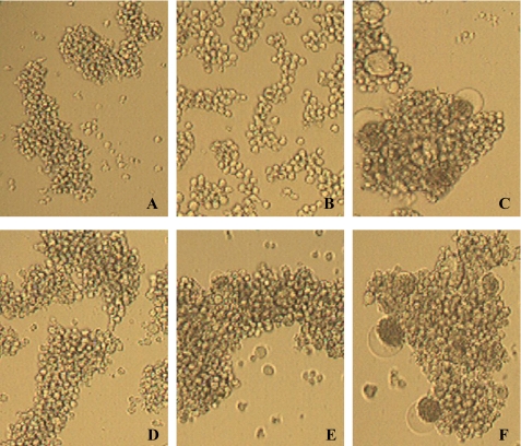
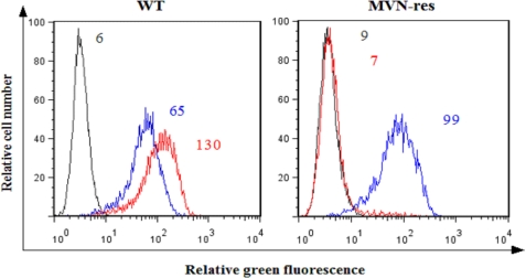
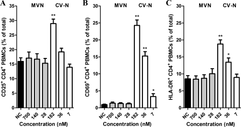
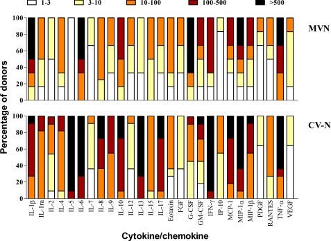
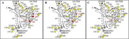
Microvirin (MVN), a recently isolated lectin from the cyanobacterium Microcystis aeruginosa PCC7806, shares 33% identity with the potent anti-human immunodeficiency virus (HIV) protein cyanovirin-N (CV-N) isolated from Nostoc ellipsosporum, and both lectins bind to similar carbohydrate structures. MVN is able to inhibit infection by a wide variety of HIV-1 laboratory-adapted strains and clinical isolates of different tropisms and subtypes in peripheral blood mononuclear cells. MVN also inhibits syncytium formation between persistently HIV-1-infected T cells and uninfected CD4(+) T cells and inhibits DC-SIGN-mediated HIV-1 binding and transmission to CD4(+) T cells. Long term passaging of HIV-1 exposed to dose-escalating concentrations of MVN resulted in the selection of a mutant virus with four deleted high mannose-type glycans in the envelope gp120. The MVN-resistant virus was still highly sensitive to various other carbohydrate binding lectins (e.g. CV-N, HHA, GNA, and UDA) but not anymore to the carbohydrate-specific 2G12 monoclonal antibody. Importantly, MVN is more than 50-fold less cytotoxic than CV-N. Also in sharp contrast to CV-N, MVN did not increase the level of the activation markers CD25, CD69, and HLA-DR in CD4(+) T lymphocytes, and subsequently, MVN did not enhance viral replication in pretreated peripheral blood mononuclear cells. Therefore, MVN may qualify as a useful lectin for potential microbicidal use based on its broad and potent antiviral activity and virtual lack of any stimulatory properties and cellular toxicity.


Figures

FIGURE 1.
FIGURE 1.
MVN inhibits giant cell formation. Shown are light microscopic pictures of the following cell cultures: SupT1 cells (A); HUT-78 cells persistently infected with HIV-1 IIIB (B); co-culture of SupT1 cells and HUT-78/IIIB cells (C); and co-culture of SupT1 cells and HUT-78/IIIB cells in the presence of MVN at 140 nm (D), 28 nm (E), and 6 nm (F).
FIGURE 2.
FIGURE 2.
No staining of 2G12 mAb on MVN-res-infected MT-4 cells. MT-4 cells were infected with WT (left) and MVN-res (right) NL4.3, and after 3–4 days, an incubation was done with the 2G12 mAb (red histograms) and the b12 mAb (blue histograms), followed by a staining with rabbit anti-human IgG-FITC. The MFI values of the background fluorescence (gray histograms) for the 2G12 mAb (red histograms) and for the b12 mAb (blue histograms) are indicated.
FIGURE 3.
FIGURE 3.
Inhibition of the binding of 2G12 mAb to HIV-1 NL4.3-infected MT-4 cells by MVN and CV-N. MT-4 cells infected with HIV-1 strain NL4.3 were incubated with 2G12 mAb in the absence (green histograms) or presence of various concentrations of MVN (red histograms, top) and CV-N (blue histograms, bottom). The light gray histograms show the background fluorescence. The MFI values of the different incubation conditions are indicated in each histogram.
FIGURE 4.
FIGURE 4.
MVN does not increase the expression of cellular activation markers. PBMCs were cultured in the presence of varying concentrations of the lectins MVN and CV-N and incubated at 37 °C. At day 3, the PBMCs were analyzed by flow cytometry for their expression of cellular activation markers with phycoerythrin-conjugated anti-CD25 (A), anti-CD69 (B), or anti-HLA-DR (C) mAbs in combination with FITC-conjugated anti-CD4 mAb. Data represent mean percentage ± S.E. for 4–16 independent experiments. *, p < 0.05; **, p < 0.0001 for comparison with untreated PBMCs (Student t test).
FIGURE 5.
FIGURE 5.
Induction of cytokines/chemokines by MVN. PBMCs from healthy donors were incubated for 72 h with medium only or MVN or CV-N at 140 and 182 nm, respectively. Supernatants were collected, and cytokine levels were measured by the Bio-Plex array system. The -fold increase values of the cytokine concentrations in the supernatant of stimulated PBMCs with respect to the concentrations in the supernatant of untreated PBMCs were determined from six and 11 different donors for MVN and CV-N, respectively. The -fold increase values are divided into subgroups: 1–3-fold increase (white squares), 3–10-fold increase (yellow squares), 10–100-fold increase (orange squares), 100–500-fold increase (dark red squares), and >500-fold increase (black squares). The amount of -fold increase values for each cytokine is given as a percentage in the total amount of donors (y axis).
FIGURE 6.
FIGURE 6.
Susceptibility of MVN-pretreated PBMCs for R5 HIV-1 infection. Freshly isolated PBMCs from two donors (A and B) were pretreated for 24 h with MVN, CV-N, HHA, and PHA and then washed and infected with the R5 HIV-1 BaL strain for 7 days. No compound was added during the infection, and viral replication was measured by a p24 Ag ELISA in the collected supernatants.
FIGURE 7.
FIGURE 7.
Ribbon diagrams showing the N-glycosylation site mutations (red circles) in gp120 of the MVN-res HIV-1 (A), the CV-N-res HIV-1 (B), and the 2G12-res HIV-1 (C). The 24 putative glycosylation sites are represented by colored circles and their accompanying amino acid number. The red circles indicate the deleted N-glycosylation sites that appear under MVN (A), CV-N (B), and 2G12 mAb (C) pressure in NL4.3 HIV-1.

References

    1. Leonard C. K., Spellman M. W., Riddle L., Harris R. J., Thomas J. N., Gregory T. J. (1990) J. Biol. Chem. 265, 10373–10382 - PubMed
    1. Kwong P. D., Wyatt R., Robinson J., Sweet R. W., Sodroski J., Hendrickson W. A. (1998) Nature 393, 648–659 - PMC - PubMed
    1. Wyatt R., Kwong P. D., Desjardins E., Sweet R. W., Robinson J., Hendrickson W. A., Sodroski J. G. (1998) Nature 393, 705–711 - PubMed
    1. O'Keefe B. R., Vojdani F., Buffa V., Shattock R. J., Montefiori D. C., Bakke J., Mirsalis J., d'Andrea A. L., Hume S. D., Bratcher B., Saucedo C. J., McMahon J. B., Pogue G. P., Palmer K. E. (2009) Proc. Natl. Acad. Sci. U.S.A. 106, 6099–6104 - PMC - PubMed
    1. Balzarini J. (2007) Nat. Rev. Microbiol. 5, 583–597 - PMC - PubMed

Publication types

MeSH terms