. 2010 Aug 6;285(32):24892-903.
doi: 10.1074/jbc.M110.147843. Epub 2010 May 27.

Structural and enzymatic insights into the ATP binding and autophosphorylation mechanism of a sensor histidine kinase

Affiliations

Structural and enzymatic insights into the ATP binding and autophosphorylation mechanism of a sensor histidine kinase

Felipe Trajtenberg et al. J Biol Chem. .

Abstract

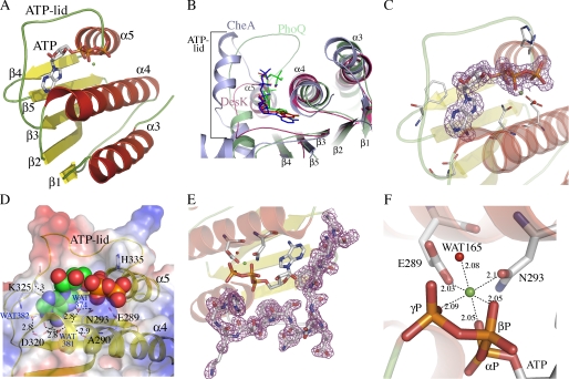
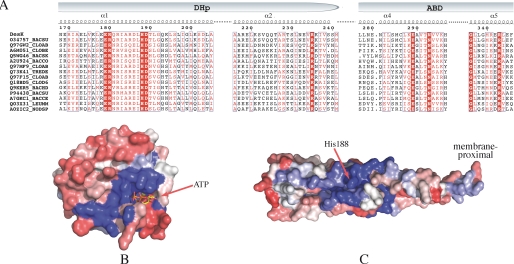
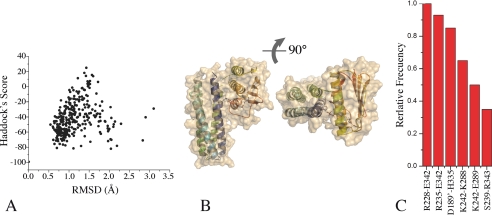
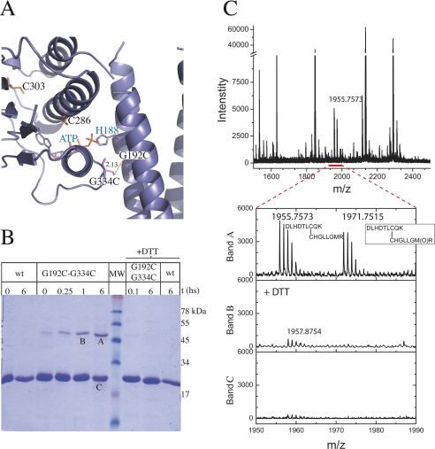
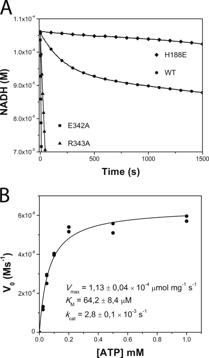
DesK is a sensor histidine kinase (HK) that allows Bacillus subtilis to respond to cold shock, triggering the adaptation of membrane fluidity via transcriptional control of a fatty acid desaturase. It belongs to the HK family HPK7, which includes the nitrogen metabolism regulators NarX/Q and the antibiotic sensor LiaS among other important sensor kinases. Structural information on different HK families is still scarce and several questions remain, particularly concerning the molecular features that determine HK specificity during its catalytic autophosphorylation and subsequent response-regulator phosphotransfer reactions. To analyze the ATP-binding features of HPK7 HKs and dissect their mechanism of autophosphorylation at the molecular level, we have studied DesK in complex with ATP using high resolution structural approaches in combination with biochemical studies. We report the first crystal structure of an HK in complex with its natural nucleotidic substrate. The general fold of the ATP-binding domain of DesK is conserved, compared with well studied members of other families. Yet, DesK displays a far more compact structure at the ATP-binding pocket: the ATP lid loop is much shorter with no secondary structural organization and becomes ordered upon ATP loading. Sequence conservation mapping onto the molecular surface, semi-flexible protein-protein docking simulations, and structure-based point mutagenesis allow us to propose a specific domain-domain geometry during autophosphorylation catalysis. Supporting our hypotheses, we have been able to trap an autophosphorylating intermediate state, by protein engineering at the predicted domain-domain interaction surface.


Figures

FIGURE 1.
FIGURE 1.
Structure of the ATP-binding domain of DesK and details of its nucleotide-binding pocket. A, schematic representation showing topology and secondary structure elements. α-Helices are colored red, β-strands are yellow, and loops are green. B, superposition of DesKABD (red tones), PhoQ (green tones), and CheA (blue tones) showing the exposure of the corresponding bound nucleotides to the solvent. Note the absence of secondary structure elements in the ATP lid of DesK. A surface representation of these differences is detailed in supplemental Fig. S2. C, 2mFobs − DFcalc electron density map contoured at 1σ, displayed around the ATP molecule, highlighting clear signal for the entire nucleotide. D, solvent-accessible surface representation of DesKABD, with the ATP moiety depicted as van der Waals balls. The surface is colored according to the mapping of electrostatic potential (red = negative, blue = positive). Key protein residues for ATP binding are highlighted in stick representation and explained in the text. The buried water H-bond network is also shown. Numbers correspond to distances in Å. E, 2mFobs − DFcalc map contoured at 1 σ, showing clear electron density for the entire ATP lid loop, ordered in the presence of ATP. F, close-up of the Mg2+ coordination site. Oxygens from the three ATP phosphates, residues Glu289 and Asn293, and water Wat165, are seen closing the octahedral coordination shell of the cation.
FIGURE 2.
FIGURE 2.
Sequence alignments and conservation patterns of the HisKA_3 subfamily. A, selected region of a reduced multiple sequence alignment with sequences from the “HisKA_3” Pfam subfamily. Identical residues are shown in bold white on a red background; conserved residues are boxed in red. Pfam IDs are indicated on the left followed by a species code: BACSU, Bacillus subtilis; CLOAB, Clostridium acetobutylicum; CLOBE, Clostridium beijerinckii (strain ncimb 8052); BACSK, Bacillus clausii (ksm-k16); BACCO, Bacillus coagulans (36d1); TREDE, Treponema denticola; CLOD6, Clostridium difficile (630); BACHD, Bacillus halodurans; BACCE, Bacillus cereus cytotoxis (nvh 391–98); LEUMM, Leuconostoc mesenteroides mesenteroides (atcc 8293/ncdo 523); and NODSP, Nodularia spumigena (ccy 9414). B, sequence conservation/variability mapped onto the molecular surface of the ATP-binding domain of DesKC. Color coding is a range from red (variable) to blue (conserved) through an intermediate white. C, same as B, mapped onto the surface of the DHp domain of DesKC.
FIGURE 3.
FIGURE 3.
Distance-restrained docking of DesKC ABD and DHp domains. A, the global Haddock score is plotted as a function of r.m.s.d. (calculated with respect to the best scoring model). Note that the best scoring models cluster at <1.5 Å r.m.s.d. B, three-dimensional structure of a representative high-scoring docked model of DesKC in the autophosphorylation state, in two orthogonal views. The solvent-accessible surface is rendered transparent to distinguish the relative organization of the domain-domain configuration (secondary structure elements are highlighted). Residues His188 and ATP are shown as sticks. C, relative frequency of interacting residue pairs in the best 10 docked structures. The histogram is limited to >30% frequencies. Engaged residues are listed, distinguishing with a single prime those belonging to the second protomer within the dimer.
FIGURE 4.
FIGURE 4.
Disulfide cross-linking assay. A, theoretical model of the mutated G192C,G334C protein in the autophosphorylation conformation, according to domain-domain docking results. Cysteine residues are shown as sticks. Predicted distance between the engineered cysteines, compatible with disulfide bond formation, is marked. B, SDS-PAGE of the cross-linking reaction. wt: wild-type DesKC used as control; G192C,G334C: DesKC double mutant with engineered cysteines (predicted molecular mass of the covalent dimer species: 50 kDa); +DTT: incubation with 0.1 m DTT. MW: molecular weight markers (corresponding masses are depicted on the right in kilodaltons). Air or air + DTT incubations were allowed to proceed for t hours. Note the DTT-induced reversion of the dimeric species to monomers. Bands A, B, and C were subsequently excised from the gel and subjected to mass spectrometry analyses. C, mass spectra of tryptic peptides derived from bands A, B, and C, showing coincidence of experimental and predicted m/z data. The spectrum segment, including peptides containing the expected disulfide bond (detailed in the inset), is enlarged. Met340 is shown to be in the reduced and oxidized forms (m/z 1955.7573 and 1971.7515, respectively).
FIGURE 5.
FIGURE 5.
Kinetic characterization of DesKC autophosphorylation. A, temporal course of ATP consumption for wild-type DesKC (WT, ○) and DesKC mutants E342A (■), R343A (▴), and H188E (autophosphorylation inactive, ♦). H188E shows a basal ATPase activity corresponding to the intrinsic activity of the ABD. Wild-type DesKC displays a biphasic behavior, with an initially exponential phase followed by a second linear regime. Note the high ATP consumption rates of E342A and R343A, which showed no biphasic pattern, even in extended incubation times until NADH depletion (not shown in this plot). B, ATP dependence of wt DesKC autophosphorylation initial velocities. Pure DesKC (22 μm) was used. kcat was calculated considering the total concentration of monomer.

Similar articles

Cited by

References

    1. Plowman G. D., Sudarsanam S., Bingham J., Whyte D., Hunter T. (1999) Proc. Natl. Acad. Sci. U.S.A. 96, 13603–13610 - PMC - PubMed
    1. Wolanin P. M., Thomason P. A., Stock J. B. (2002) Genome Biol. 3, REVIEWS3013
    1. Machius M., Chuang J. L., Wynn R. M., Tomchick D. R., Chuang D. T. (2001) Proc. Natl. Acad. Sci. U.S.A. 98, 11218–11223 - PMC - PubMed
    1. Hsing W., Russo F. D., Bernd K. K., Silhavy T. J. (1998) J. Bacteriol. 180, 4538–4546 - PMC - PubMed
    1. Laub M. T., Goulian M. (2007) Annu. Rev. Genet 41, 121–145 - PubMed

Publication types

Associated data

LinkOut - more resources