. 2010 Aug 6;285(32):24313-23.
doi: 10.1074/jbc.M110.118398. Epub 2010 May 27.

cAMP-regulated protein lysine acetylases in mycobacteria

Affiliations

cAMP-regulated protein lysine acetylases in mycobacteria

Subhalaxmi Nambi et al. J Biol Chem. .

Abstract

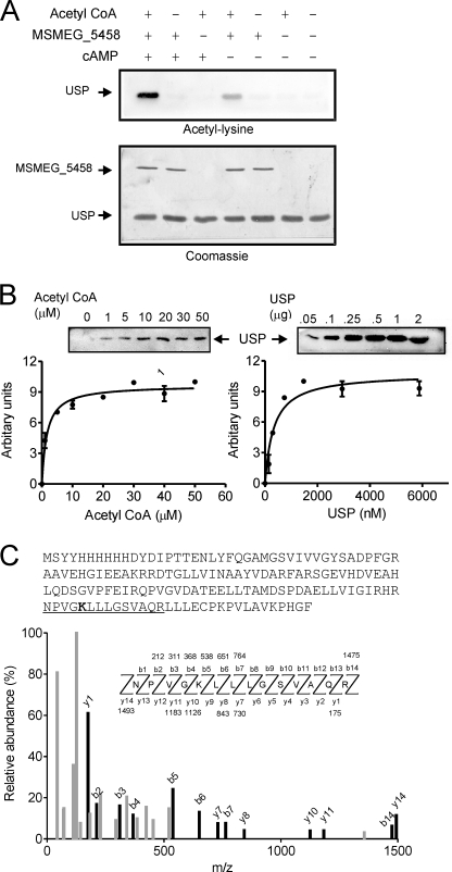
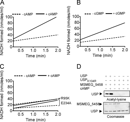
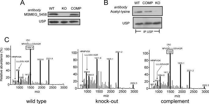
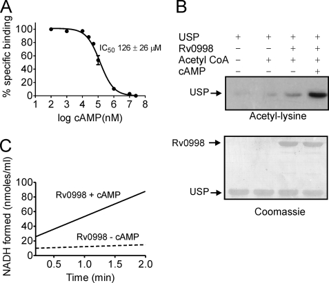
Cyclic AMP synthesized by Mycobacterium tuberculosis has been shown to play a role in pathogenesis. However, the high levels of intracellular cAMP found in both pathogenic and non-pathogenic mycobacteria suggest that additional and important biological processes are regulated by cAMP in these organisms. We describe here the biochemical characterization of novel cAMP-binding proteins in M. smegmatis and M. tuberculosis (MSMEG_5458 and Rv0998, respectively) that contain a cyclic nucleotide binding domain fused to a domain that shows similarity to the GNAT family of acetyltransferases. We detect protein lysine acetylation in mycobacteria and identify a universal stress protein (USP) as a substrate of MSMEG_5458. Acetylation of a lysine residue in USP is regulated by cAMP, and using a strain deleted for MSMEG_5458, we show that USP is indeed an in vivo substrate for MSMEG_5458. The Rv0998 protein shows a strict cAMP-dependent acetylation of USP, despite a lower affinity for cAMP than MSMEG_5458. Thus, this report not only represents the first demonstration of protein lysine acetylation in mycobacteria but also describes a unique functional interplay between a cyclic nucleotide binding domain and a protein acetyltransferase.


Figures

FIGURE 1.
FIGURE 1.
Sequence alignment of cNMP binding and acetyltransferase domains. A, multiple sequence alignment of the cNMP binding domains of Rv0998, MSMEG_5458, regulatory subunits of PKA (RIα, RIIα, and RIIβ), exchange protein activated by cAMP, hyperpolarization-activated cyclic nucleotide, and CRP from M. tuberculosis. The Protein Data Bank codes of structurally characterized proteins are shown. Asterisks represent important residues that are present in cNMP binding domains, which are discussed in the text. The black arrow represents the arginine (Arg-95 in MSMEG_5458) that is important for cAMP binding. Inset, a diagrammatic representation of the domain organization of MSMEG_5458/Rv0998. B, phylogenetic clustering of the cNMP binding domains in Rv0998 and MSMEG_5458. The gi numbers (supplemental Table S2, and in some cases, common names along with gi numbers) are shown, and A or B following the gi numbers identifies the first (N-terminal) or the second cNMP binding domain seen in the full-length protein sequences. PKA:A, cAMP binding domains in the regulatory subunits of PKA (A domain); GEF, guanine nucleotide exchange factor; PKA:B, cAMP binding domains in the regulatory subunits of PKA (B domain); PKG:A, cGMP binding domains in cGMP-dependent protein kinases (A domain); PKG:B, cGMP binding domains in cGMP-dependent protein kinases (B domain); CNG, cyclic nucleotide gated channels; NTE, neuropathy target esterase (also known as lysophospholipase NTE). Black dots indicate MSMEG_5458 and Rv0998. C, multiple sequence alignment of the acetyltransferase domain of Rv0998, MSMEG_5458, human GCN5 acetyltransferase, yeast histone acetyltransferase and tetrahymena GCN5 acetyltransferase. The Protein Data Bank codes of structurally characterized proteins are shown in the alignment. Asterisks represent important residues that are present in cNMP binding domains, which are discussed in the text. The black arrow identifies the glutamate residue important for acetyltransferase activity.
FIGURE 2.
FIGURE 2.
cAMP binding to MSMEG_5458. A, displacement of [3H]cAMP from MSMEG_5458 (∼50 nm) by unlabeled cyclic nucleotides (10 μm). Values represent the mean ± S.E.; n = 3. B, displacement of [3H]cAMP (∼50 nm) from MSMEG_5458 with increasing concentrations of the indicated ligands; n = 3. Values represent the mean ± S.E. C, binding of [3H]cAMP (∼50 nm) to MSMEG_5458R95K. Values shown represent the mean ± S.E. of duplicate determinations in experiments repeated thrice.
FIGURE 3.
FIGURE 3.
Protein acetylation in mycobacteria and identification of USP as an interacting partner of MSMEG_5458. A, whole cell lysates (50 μg) prepared from cultures of M. tuberculosis H37Rv, M. bovis Bacillus Calmette-Guérin, and M. smegmatis were separated by SDS-gel electrophoresis and Western blotting performed with acetyl-lysine antibodies in the presence or absence of acetylated bovine serum albumin. B, GST or GST-MSMEG_5458 bound to glutathione beads were incubated with cytosolic fractions prepared from M. smegmatis, in the presence or absence of 10 μm cAMP. Interacting proteins were resolved by SDS-PAGE and visualized by staining with Coomassie Brilliant Blue. One protein (*) was analyzed by mass spectrometry and identified to be MSMEG_4207 (USP). C, samples obtained after pulldown experiments under the conditions indicated were analyzed by Western blotting with acetyl-lysine antibodies. Experiments were repeated thrice.
FIGURE 4.
FIGURE 4.
Acetylation of USP and identification of the site of acetylation. A, USP (2 μg) was incubated alone, or in the presence of MSMEG_5458 (100 ng), 10 μm cAMP, and acetyl-CoA (30 μm) as indicated at 25 °C for 10 min, followed by Western blotting with acetyl-lysine antibodies (upper panel). Following blotting, the membrane was stained with Coomassie Brilliant Blue (lower panel). B, acetylation of USP was monitored in a Western blot based assay (“Experimental Procedures”). Left panel, assays performed in the presence of varying concentrations of acetyl-CoA with a fixed concentration of USP (25 μm). Right panel, assays performed in the presence of varying concentrations of USP and a fixed concentration of acetyl-CoA (30 μm). All assays were incubated at 25 °C for 10 min. Insets show representative blots obtained from the assays. Values represent the mean ± S.E. of assays performed thrice. C, tandem mass (MS/MS) spectrum of a tryptic peptide of mass/charge ratio (m/z) 1493.8 obtained from acetylated USP. Singly charged fragment ions marked in the spectrum in black represent peptide bond cleavage resulting in sequence information recorded from both the N and C termini (b- and y-type ions, respectively). This spectrum matched that of the peptide (underlined) in USP (sequence of purified protein showed as an inset), with mass shift in the b5 and y10 ions corresponding to acetylation at the lysine residue (bold in inset).
FIGURE 5.
FIGURE 5.
Initial rates of the acetyltransferase activity of MSMEG_5458. The acetyltransferase activities of wild type (A and B) and mutant MSMEG_5458 proteins (C) were measured using the coupled assay. MSMEG_5458 or mutant proteins (1 μg) were assayed in the presence of 30 μm acetyl-CoA and 50 μm USP. The initial rate of formation of NADH is shown, after subtracting the change in absorbance at 340 nm that is seen in assays performed in the absence of the enzymes, which was usually ∼1% of the change seen in the presence of enzyme. D, USPK104R (50 μm) was used as substrate for the acetyltransferase activity, in the presence or absence of cAMP (10 μm). Samples were subjected to Western blotting using acetyl-lysine antibodies, and either wild-type USP or USPK104R. Data shown are a representative of assays performed thrice.
FIGURE 6.
FIGURE 6.
USP is a substrate of MSMEG_5458 in vivo. A, Western blot analysis of whole cell lysates prepared from wild type (WT), ΔMSMEG_5458 (KO), and ΔMSMEG_5458 complemented with wild-type MSMEG_5458 (COMP) strains using polyclonal MSMEG_5458 and USP antibodies. B, whole cell lysates from WT, KO, and COMP were immunoprecipitated with USP antibody and immunoprecipitates subjected to Western blot analysis with acetyl-lysine antibody followed by normalization with USP polyclonal antibodies. C, MALDI mass spectra of in-gel tryptic digests of immunoprecipitated USP obtained from WT, KO, and COMP cells. The peaks in black represent peptides from USP. The 1493.8-Da peptide (represented as a peak marked with a box) corresponds to the acetylated peptide and is present in USP from WT and COMP strains but absent in the KO strain.
FIGURE 7.
FIGURE 7.
Biochemical properties of Rv0998. A, displacement of [3H]cAMP (∼50 nm) from Rv0998 (5 μg) with increasing concentrations of unlabeled cAMP. Values represent the mean ± S.E. with n = 3. B, acetylation of USP by Rv0998 was monitored by Western blot analysis with acetyl-lysine antibodies performed in the presence or absence of cAMP (1 mm, upper panel) followed by staining of the blot with Coomassie Brilliant Blue (lower panel). C, acetyltransferase activity of Rv0998 using the coupled assay, in the absence or presence of 1 mm cAMP. Data are representative of assays performed thrice.

Similar articles

Cited by

References

    1. Barzu O., Danchin A. (1994) Prog. Nucleic Acids Res. Mol. Biol. 49, 241–283 - PubMed
    1. Linder J. U., Schultz J. E. (2003) Cell Signal. 15, 1081–1089 - PubMed
    1. Shenoy A. R., Visweswariah S. S. (2006) FEBS Lett. 580, 3344–3352 - PubMed
    1. Shenoy A. R., Sreenath N., Podobnik M., Kovacevic M., Visweswariah S. S. (2005) Biochemistry 44, 15695–15704 - PubMed
    1. Podobnik M., Tyagi R., Matange N., Dermol U., Gupta A. K., Mattoo R., Seshadri K., Visweswariah S. S. (2009) J. Biol. Chem. 284, 32846–32857 - PMC - PubMed

Publication types

LinkOut - more resources