

Location, location, location: compartmentalization of early events in leukotriene biosynthesis
- PMID: 20507998
- PMCID: PMC2919072
- DOI: 10.1074/jbc.R110.125880
Location, location, location: compartmentalization of early events in leukotriene biosynthesis
Erratum in
- J Biol Chem. 2010 Dec 3;285(49):38740
Abstract
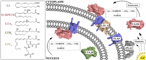
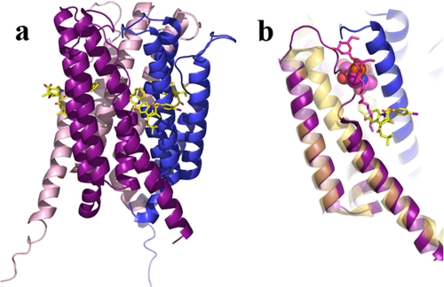
Leukotrienes (LTs), derived from arachidonic acid (AA) released from the membrane by the action of phospholipase A(2), are potent lipid mediators of the inflammatory response. In 1983, Dahlén et al. demonstrated that LTC(4), LTD(4), and LTE(4) mediate antigen-induced constriction of bronchi in tissue obtained from subjects with asthma (Dahlén, S. E., Hansson, G., Hedqvist, P., Björck, T., Granström, E., and Dahlén, B. (1983) Proc. Natl. Acad. Sci. U.S.A. 80, 1712-1716). Over the last 25+ years, substantial progress has been made in understanding how LTs exert their effects, and a broader appreciation for the numerous biological processes they mediate has emerged. LT biosynthesis is initiated by the action of 5-lipoxygenase (5-LOX), which catalyzes the transformation of AA to LTA(4) in a two-step reaction. Ca(2+) targets 5-LOX to the nuclear membrane, where it co-localizes with the 5-LOX-activating protein FLAP and, when present, the downstream enzyme LTC(4) synthase, both transmembrane proteins. Crystal structures of the AA-metabolizing LOXs, LTC(4) synthase, and FLAP combined with biochemical data provide a framework for understanding how subcellular organizations optimize the biosynthesis of these labile hydrophobic signaling compounds, which must navigate pathways that include both membrane and soluble enzymes. The insights these structures afford and the questions they engender are discussed in this minireview.
Figures


Similar articles
-
Structures and mechanisms of enzymes in the leukotriene cascade.Biochimie. 2010 Jun;92(6):676-81. doi: 10.1016/j.biochi.2010.01.010. Epub 2010 Jan 22. Biochimie. 2010. PMID: 20097252 Review.
-
Increased expression of leukotriene C4 synthase and predominant formation of cysteinyl-leukotrienes in human abdominal aortic aneurysm.Proc Natl Acad Sci U S A. 2010 Dec 7;107(49):21093-7. doi: 10.1073/pnas.1015166107. Epub 2010 Nov 15. Proc Natl Acad Sci U S A. 2010. PMID: 21078989 Free PMC article.
-
Untangling the web of 5-lipoxygenase-derived products from a molecular and structural perspective: The battle between pro- and anti-inflammatory lipid mediators.Biochem Pharmacol. 2021 Nov;193:114759. doi: 10.1016/j.bcp.2021.114759. Epub 2021 Sep 3. Biochem Pharmacol. 2021. PMID: 34487716 Free PMC article. Review.
-
5-Lipoxygenase metabolic contributions to NSAID-induced organ toxicity.Adv Ther. 2012 Feb;29(2):79-98. doi: 10.1007/s12325-011-0100-7. Epub 2012 Feb 7. Adv Ther. 2012. PMID: 22351432 Review.
-
Transcellular biosynthesis contributes to the production of leukotrienes during inflammatory responses in vivo.J Clin Invest. 2002 May;109(10):1373-80. doi: 10.1172/JCI14869. J Clin Invest. 2002. PMID: 12021253 Free PMC article.
Cited by
-
Impact of Androgens on Inflammation-Related Lipid Mediator Biosynthesis in Innate Immune Cells.Front Immunol. 2020 Jun 30;11:1356. doi: 10.3389/fimmu.2020.01356. eCollection 2020. Front Immunol. 2020. PMID: 32714332 Free PMC article. Review.
-
Biochemical basis of asthma therapy.J Biol Chem. 2011 Sep 23;286(38):32899-905. doi: 10.1074/jbc.R110.206466. Epub 2011 Jul 28. J Biol Chem. 2011. PMID: 21799015 Free PMC article. Review.
-
Eicosanoid storm in infection and inflammation.Nat Rev Immunol. 2015 Aug;15(8):511-23. doi: 10.1038/nri3859. Epub 2015 Jul 3. Nat Rev Immunol. 2015. PMID: 26139350 Free PMC article. Review.
-
Fluctuations of an exposed π-helix involved in lipoxygenase substrate recognition.Biochemistry. 2014 Aug 12;53(31):5102-10. doi: 10.1021/bi500768c. Epub 2014 Jul 29. Biochemistry. 2014. PMID: 25036469 Free PMC article.
-
CD36 protein is involved in store-operated calcium flux, phospholipase A2 activation, and production of prostaglandin E2.J Biol Chem. 2011 May 20;286(20):17785-95. doi: 10.1074/jbc.M111.232975. Epub 2011 Mar 31. J Biol Chem. 2011. PMID: 21454644 Free PMC article.
References
-
- Brash A. R. (1999) J. Biol. Chem. 274, 23679–23682 - PubMed
-
- Kuhn H., Thiele B. J. (1999) FEBS Lett. 449, 7–11 - PubMed
-
- Noguchi M., Miyano M., Matsumoto T., Noma M. (1994) Biochim. Biophys. Acta 1215, 300–306 - PubMed
-
- Hill E., Maclouf J., Murphy R. C., Henson P. M. (1992) J. Biol. Chem. 267, 22048–22053 - PubMed
-
- Boyington J. C., Gaffney B. J., Amzel L. M. (1993) Science 260, 1482–1486 - PubMed
Publication types
MeSH terms
Substances
LinkOut - more resources
Full Text Sources
Miscellaneous
