

2009 pandemic influenza A (H1N1): pathology and pathogenesis of 100 fatal cases in the United States
- PMID: 20508031
- PMCID: PMC2893660
- DOI: 10.2353/ajpath.2010.100115
2009 pandemic influenza A (H1N1): pathology and pathogenesis of 100 fatal cases in the United States
Abstract
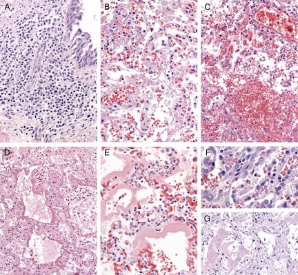
In the spring of 2009, a novel influenza A (H1N1) virus emerged in North America and spread worldwide to cause the first influenza pandemic since 1968. During the first 4 months, over 500 deaths in the United States had been associated with confirmed 2009 pandemic influenza A (H1N1) [2009 H1N1] virus infection. Pathological evaluation of respiratory specimens from initial influenza-associated deaths suggested marked differences in viral tropism and tissue damage compared with seasonal influenza and prompted further investigation. Available autopsy tissue samples were obtained from 100 US deaths with laboratory-confirmed 2009 H1N1 virus infection. Demographic and clinical data of these case-patients were collected, and the tissues were evaluated by multiple laboratory methods, including histopathological evaluation, special stains, molecular and immunohistochemical assays, viral culture, and electron microscopy. The most prominent histopathological feature observed was diffuse alveolar damage in the lung in all case-patients examined. Alveolar lining cells, including type I and type II pneumocytes, were the primary infected cells. Bacterial co-infections were identified in >25% of the case-patients. Viral pneumonia and immunolocalization of viral antigen in association with diffuse alveolar damage are prominent features of infection with 2009 pandemic influenza A (H1N1) virus. Underlying medical conditions and bacterial co-infections contributed to the fatal outcome of this infection. More studies are needed to understand the multifactorial pathogenesis of this infection.
Figures


Comment in
-
The 2009 H1N1 pandemic adds to our knowledge of influenza pathogenesis.Am J Pathol. 2010 Jul;177(1):13-4. doi: 10.2353/ajpath.2010.100351. Epub 2010 May 20. Am J Pathol. 2010. PMID: 20489147 Free PMC article.
References
-
- Dawood FS, Jain S, Finelli L, Shaw MW, Lindstrom S, Garten RJ, Gubareva LV, Xu X, Bridges CB, Uyeki TM. Emergence of a novel swine-origin influenza A (H1N1) virus in humans. N Engl J Med. 2009;360:2605–2615. - PubMed
-
- Echevarria-Zuno S, Mejia-Arangure JM, Mar-Obeso AJ, Grajales-Muniz C, Robles-Perez E, Gonzalez-Leon M, Ortega-Alvarez MC, Gonzalez-Bonilla C, Rascon-Pacheco RA, Borja-Aburto VH. Infection and death from influenza A H1N1 virus in Mexico: a retrospective analysis. Lancet. 2009;374:2072–2079. - PubMed
-
- Jain S, Kamimoto L, Bramley AM, Schmitz AM, Benoit SR, Louie J, Sugerman DE, Druckenmiller JK, Ritger KA, Chugh R, Jasuja S, Deutscher M, Chen S, Walker JD, Duchin JS, Lett S, Soliva S, Wells EV, Swerdlow D, Uyeki TM, Fiore AE, Olsen SJ, Fry AM, Bridges CB, Finelli L. Hospitalized patients with 2009 H1N1 influenza in the United States. April–June 2009. N Engl J Med. 2009;361:1935–1944. - PubMed
-
- Louie JK, Acosta M, Winter K, Jean C, Gavali S, Schechter R, Vugia D, Harriman K, Matyas B, Glaser CA, Samuel MC, Rosenberg J, Talarico J, Hatch D. Factors associated with death or hospitalization due to pandemic 2009 influenza A(H1N1) infection in California. JAMA. 2009;302:1896–1902. - PubMed
-
- Miller RR, 3rd, Markewitz BA, Rolfs RT, Brown SM, Dascomb KK, Grissom CK, Friedrichs MD, Mayer J, Hirshberg EL, Conklin J, Paine R, 3rd, Dean NC. Clinical findings and demographic factors associated with ICU admission in Utah due to novel 2009 influenza A (H1N1) infection. Chest. 2010;137:752–758. - PMC - PubMed
MeSH terms
LinkOut - more resources
Full Text Sources
Other Literature Sources
Medical
