. 2010 Jul 9;329(5988):182-6.
doi: 10.1126/science.1190414. Epub 2010 May 27.

Structure of the human BK channel Ca2+-activation apparatus at 3.0 A resolution

Affiliations

Structure of the human BK channel Ca2+-activation apparatus at 3.0 A resolution

Peng Yuan et al. Science. .

Abstract

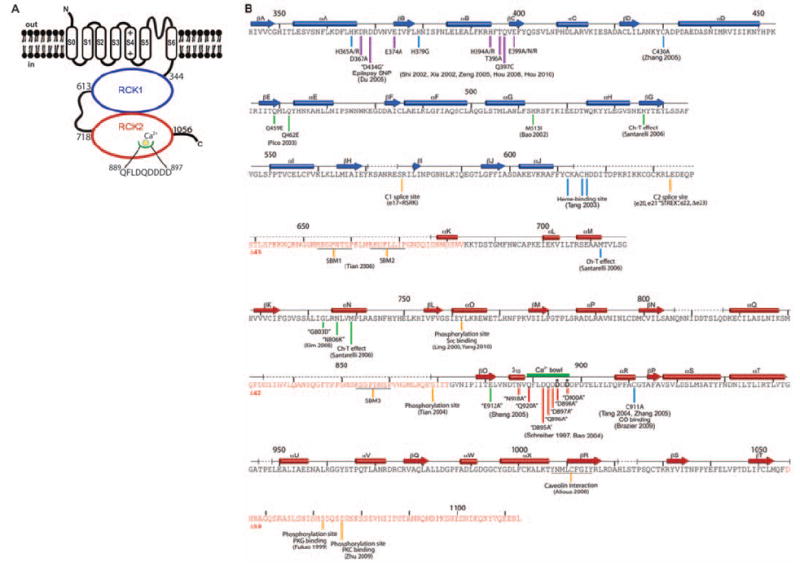
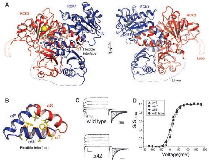
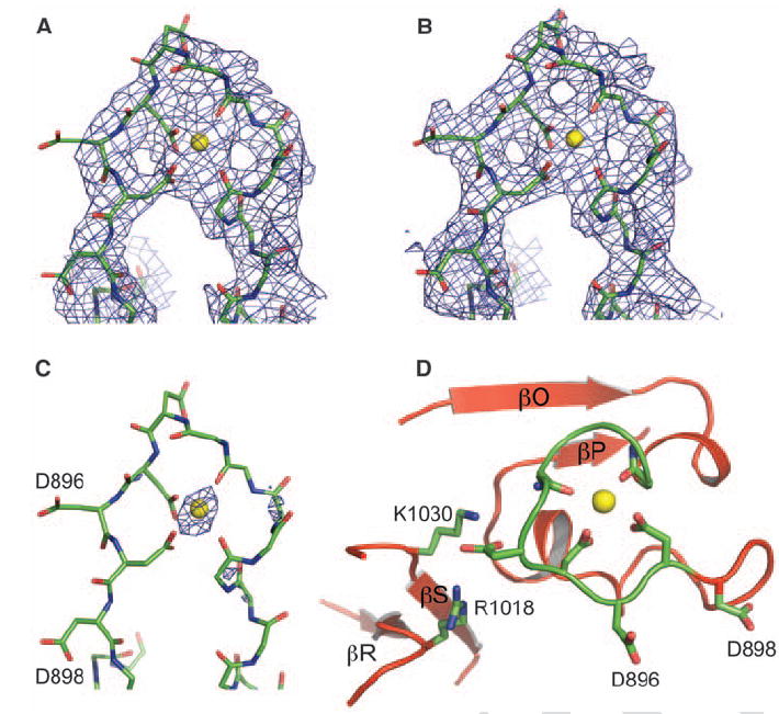
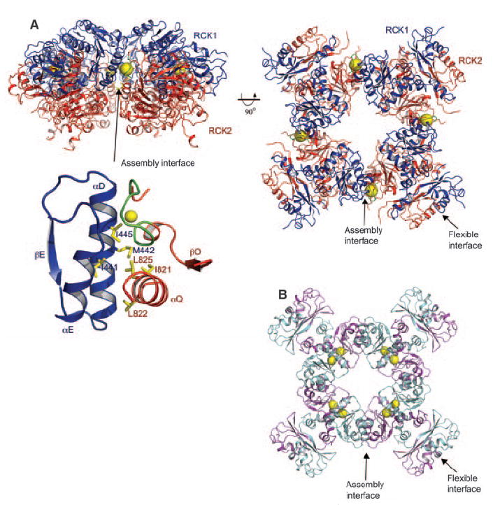
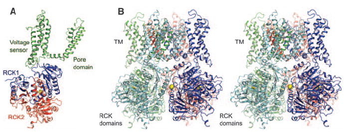
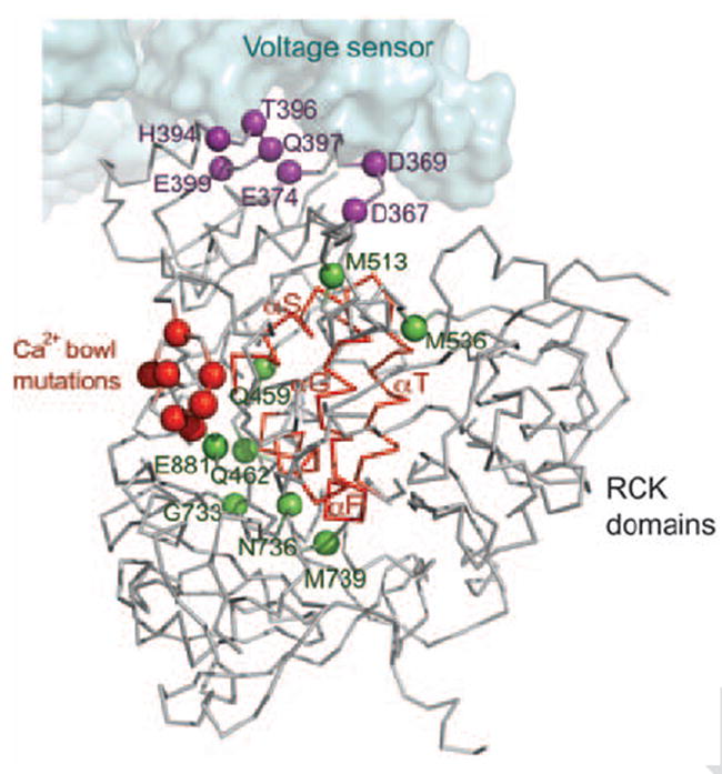
High-conductance voltage- and Ca2+-activated K+ (BK) channels encode negative feedback regulation of membrane voltage and Ca2+ signaling, playing a central role in numerous physiological processes. We determined the x-ray structure of the human BK Ca2+ gating apparatus at a resolution of 3.0 angstroms and deduced its tetrameric assembly by solving a 6 angstrom resolution structure of a Na+-activated homolog. Two tandem C-terminal regulator of K+ conductance (RCK) domains from each of four channel subunits form a 350-kilodalton gating ring at the intracellular membrane surface. A sequence of aspartic amino acids that is known as the Ca2+ bowl, and is located within the second of the tandem RCK domains, creates four Ca2+ binding sites on the outer perimeter of the gating ring at the "assembly interface" between RCK domains. Functionally important mutations cluster near the Ca2+ bowl, near the "flexible interface" between RCK domains, and on the surface of the gating ring that faces the voltage sensors. The structure suggests that the Ca2+ gating ring, in addition to regulating the pore directly, may also modulate the voltage sensor.


Figures

Fig. 1
Fig. 1
The BK channel. (A) Domain topology of BK. Domain boundaries are indicated, and the Ca2+ bowl is illustrated. (B) Secondary structure of human BK CTD. Secondary structure elements from the crystal structure are aligned to the BK sequence, showing tandem RCK domains (RCK1, blue; RCK2, red) with unresolved segments indicated by dashed lines. Deletion mutants Δ45, Δ42, and Δ58 of unresolved segments (sequences highlighted in red) were characterized functionally (Fig. 2, C and D). The Ca2+ bowl is indicated as a thick green line. Site-specific functional data are marked beneath the sequence by vertical lines with references. The complete list of corresponding references is given in table S3. The color of the lines specify the following categories: red, mutations around the Ca2+ bowl; purple, mutations facing the voltage sensor domain in our BK model (Fig. 6); green, mutations near the αF-αG/αS-αT flexible interface; yellow, splice sites, protein-protein interaction sites, and phosphorylation sites; and blue, additional sites of mutation and modification.
Fig. 2
Fig. 2
Structure of the BK CTD. (A) Ribbon representations of front and back views of the BK CTD showing RCK1 in blue and RCK2 in red. The Ca2+ bowl is colored in green, and the Ca2+ ion is shown as a yellow sphere. Large disordered segments are indicated as dashed lines. (B) Close-up view of the αF-αG/αS-αT flexible interface. Side chains of hydrophobic residues in the interface are shown as sticks. (C) Channel activation. Currents from inside-out patches are shown for wild-type BK and Δ42 deletion mutant in the presence of 10 μM Ca2+. Voltage pulses are −120 to +180 mV, ΔV = 20 mV, and a holding potential of −60 mV. (D) Normalized G-V curves for wild type and deletion mutants in 10 μM Ca2+. Δ45* was tested by means of co-expression of two separated channel segments instead of deletion from a single construct.
Fig. 3
Fig. 3
The Ca2+ bowl. (A) Experimental electron density at 3.3 Å in the Ca2+ bowl region contoured at 1.0 above the mean density, where σ is the RMSD of the density (B and C) Weighted 2fo-fc electron density at 3.0 Å after refinement contoured at 1.0 σ and 4.0 σ, respectively. The final refined model is shown as sticks, with most side chains removed for clarity. (D) Structure of the Ca2+ bowl, showing key residues coordinating the Ca2+ ion (yellow sphere).
Fig. 4
Fig. 4
The BK gating ring solution. (A) Orthogonal views of the tetrameric gating ring structure using 6.0 Å diffraction data from the homologous chicken Slo2.2 CTD. (Right) View is down the fourfold symmetry axis, with RCK1 in blue and RCK2 in red. The Ca2+ ions are shown as yellow spheres. The flexible and assembly interfaces are labeled, and a close-up view of the assembly interface is also shown. (B) Open gating ring structure from the MthK channel (PDB accession, 1LNQ), viewed down the fourfold axis.
Fig. 5
Fig. 5
A model of the BK channel. (A) Ribbon representation of a single subunit from the BK model generated by superimposing the BK gating ring and Kv paddle chimera (PDB accession, 2R9R) onto the MthK channel (PDB accession, 1LNQ). The transmembrane domain, RCK1, and RCK2 are colored in green, blue, and red, respectively. (B) Stereoview of the BK model from the side with the transmembrane domain above. Each subunit of the tetramer is colored differently.
Fig. 6
Fig. 6
The gating ring and the voltage sensor modules. Sensitive functional mutations from the CTD (colored spheres) were mapped onto a single subunit from the BK model. Three distinct groups of mutations are distinguished by using the same colors as in Fig. 1B. The Ca2+ bowl mutations are colored in red. Mutations near the αF-αG/αS-αT flexible interface (backbone highlighted in red) are colored in green, and mutations in the RCK1 domain facing the voltage sensor domain are colored in purple. The voltage sensor domain is shown as a surface representation.

Comment in

References

    1. Robitaille R, Garcia ML, Kaczorowski GJ, Charlton MP. Neuron. 1993;11:645. - PubMed
    1. Fettiplace R, Fuchs PA. Annu Rev Physiol. 1999;61:809. - PubMed
    1. Nelson MT, et al. Science. 1995;270:633. - PubMed
    1. Brenner R, et al. Nature. 2000;407:870. - PubMed
    1. Petkov GV, et al. J Physiol. 2001;537:443. - PMC - PubMed

Publication types

MeSH terms

Associated data