. 2010 May 28;328(5982):1172-6.
doi: 10.1126/science.1187532.

mTORC1-mediated cell proliferation, but not cell growth, controlled by the 4E-BPs

Affiliations

mTORC1-mediated cell proliferation, but not cell growth, controlled by the 4E-BPs

Ryan J O Dowling et al. Science. .

Abstract

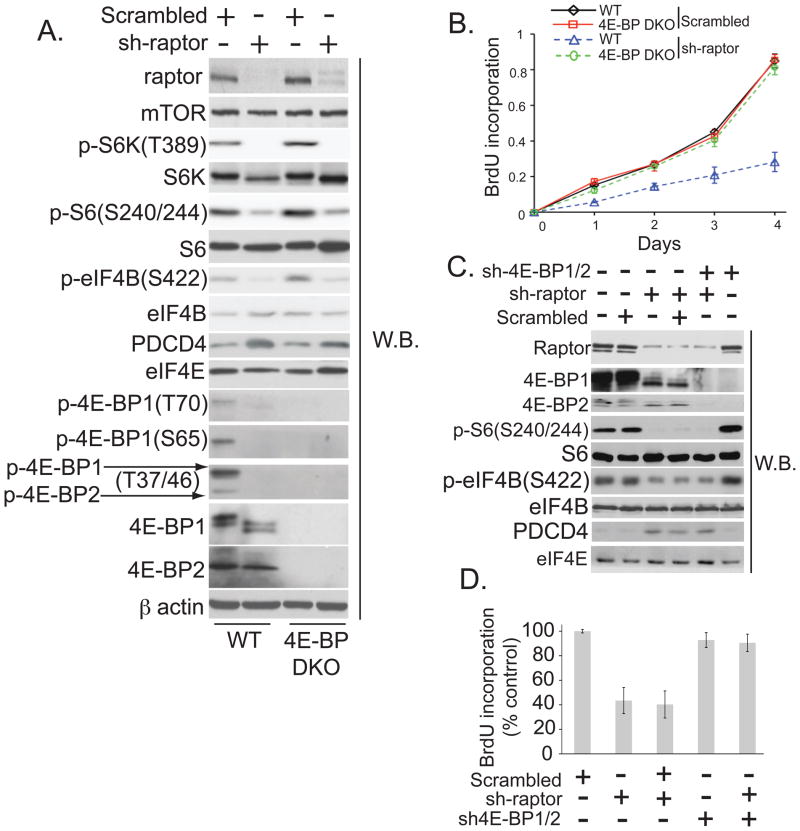
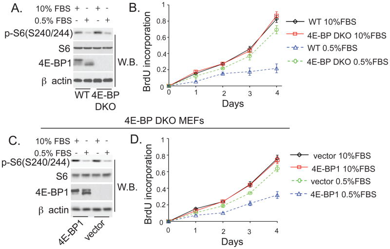
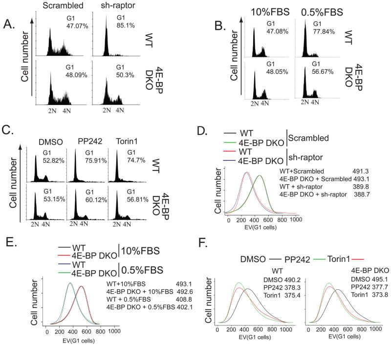
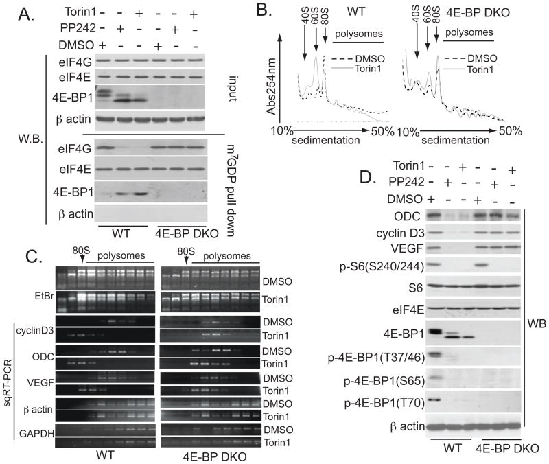
The mammalian target of rapamycin complex 1 (mTORC1) integrates mitogen and nutrient signals to control cell proliferation and cell size. Hence, mTORC1 is implicated in a large number of human diseases--including diabetes, obesity, heart disease, and cancer--that are characterized by aberrant cell growth and proliferation. Although eukaryotic translation initiation factor 4E-binding proteins (4E-BPs) are critical mediators of mTORC1 function, their precise contribution to mTORC1 signaling and the mechanisms by which they mediate mTORC1 function have remained unclear. We inhibited the mTORC1 pathway in cells lacking 4E-BPs and analyzed the effects on cell size, cell proliferation, and cell cycle progression. Although the 4E-BPs had no effect on cell size, they inhibited cell proliferation by selectively inhibiting the translation of messenger RNAs that encode proliferation-promoting proteins and proteins involved in cell cycle progression. Thus, control of cell size and cell cycle progression appear to be independent in mammalian cells, whereas in lower eukaryotes, 4E-BPs influence both cell growth and proliferation.


Figures

Fig. 1
Fig. 1. 4E-BPs mediate mTORC1’s effects on cell proliferation
(A) Levels of the indicated proteins in wild-type and 4E-BP DKO MEFs infected with a scrambled or raptor-specific shRNA (sh-raptor) were determined by Western blotting. β actin-loading control. (B) Proliferation of MEFs from (A) was determined by 5-bromo2′-deoxyuridine (BrdU) incorporation. Results represent means ± SD (n=4). (C) Western blots of HEK293T cells infected by raptor and 4E-BP1 and 2 shRNA (sh-raptor; sh-4E-BP1/2), or scrambled shRNA. eIF4E served as a loading control. D Proliferation of cells from (C) was determined by BrdU incorporation. Results represent percentage of the values obtained in dimethyl sulfoxide (DMSO)-treated cells (set to 100%) ± SD values (n=4).
Fig. 2
Fig. 2. 4E-BPs regulate cell proliferation in low-serum conditions
(A and C) Levels of the indicated proteins were determined by Western blotting in wild-type and 4E-BP DKO MEFs (A), or 4E-BP DKO MEFs infected with vector or 4E-BP1 construct (C), and maintained for 48h in 10% or 0.5% FBS. β actin served as a loading control. (B and D) Proliferation of WT and 4E-BP DKO MEFs (B) or 4E-BP DKO MEFs infected with vector or 4E-BP1 construct (D) maintained in 10% or 0.5% FBS was measured by BrdU incorporation. Results represent means ± SD (n=3).
Fig. 3
Fig. 3. 4E-BPs mediate the effects of mTORC1 signaling on cell cycle, but not on cell size
(A to C) Cell cycle distributions for WT and 4E-BP DKO MEFs infected with a control or raptor-specific (sh-raptor) shRNA (A), maintained in 10% or 0.5% FBS for 48 hours (B), or treated with 2.5 μM PP242, 250 nM Torin1, or vehicle (DMSO) for 24 hours (C) were monitored by flow cytometry. N indicates DNA content. Cell size distributions for WT and 4E-BP DKO MEFs infected with a scrambled control or raptor-specific (sh-raptor) shRNA (D), maintained in 10 or 0.5% FBS (E) or treated as in (C) with the indicated compounds (F). The size of G1 cells was determined by measuring electronic volume (EV) with flow cytometry; numbers represent mean EV values.
Fig. 4
Fig. 4. asTORi inhibit translation of mRNAs encoding proliferation-promoting proteins via 4E-BPs
(A) WT and 4E-BP DKO MEFs treated for 24 hours with 2.5 μM PP242, 250 nM Torin1, or vehicle (DMSO) were subjected to m7GDP pull-down. Amounts of the indicated proteins in the input (10%) or pull-down (25%) were determined by Western blotting. β actin served as a loading control (input) and to exclude contamination (m7GDP pull-down). (B) Absorption profiles of ribosomes from WT and 4E-BP DKO cells treated for 24 hours with 250 nM Torin1, or vehicle (DMSO). 40S and 60S denote the corresponding ribosomal subunits; 80S, monosome. (C) RNA was visualized by ethidium bromide (EtBr). Distribution of cyclin D3, VEGF, ODC, β actin, and GAPDH mRNAs was determined by semiquantitative reverse transcription polymerase chain reaction (sqRT-PCR). The RT-PCR reactions were in the linear range (fig. S7I). (D) WT and 4E-BP DKO MEFs were treated as in (A), and the amount of the indicated proteins was determined by Western blotting. β actin served as a loading control.

References

    1. Guertin DA, Sabatini DM. Cancer Cell. 2007 Jul;12:9. - PubMed
    1. Hay N, Sonenberg N. Genes Dev. 2004 Aug 15;18:1926. - PubMed
    1. Pause A, et al. Nature. 1994 Oct 27;371:762. - PubMed
    1. Choo AY, Blenis J. Cell Cycle. 2009 Feb 15;8:567. - PubMed
    1. Hara K, et al. Cell. 2002 Jul 26;110:177. - PubMed

Publication types

MeSH terms