Review
. 2010 Jun;74(2):229-49.
doi: 10.1128/MMBR.00043-09.

Gliding motility revisited: how do the myxobacteria move without flagella?

Affiliations
Review

Gliding motility revisited: how do the myxobacteria move without flagella?

Emilia M F Mauriello et al. Microbiol Mol Biol Rev. 2010 Jun.

Abstract

In bacteria, motility is important for a wide variety of biological functions such as virulence, fruiting body formation, and biofilm formation. While most bacteria move by using specialized appendages, usually external or periplasmic flagella, some bacteria use other mechanisms for their movements that are less well characterized. These mechanisms do not always exhibit obvious motility structures. Myxococcus xanthus is a motile bacterium that does not produce flagella but glides slowly over solid surfaces. How M. xanthus moves has remained a puzzle that has challenged microbiologists for over 50 years. Fortunately, recent advances in the analysis of motility mutants, bioinformatics, and protein localization have revealed likely mechanisms for the two M. xanthus motility systems. These results are summarized in this review.


Figures

FIG. 1.
FIG. 1.
Life cycle of M. xanthus. (Vegetative growth) On a solid surface with soluble nutrients, groups of M. xanthus cells (swarms) grow, divide, and move outward. On a solid surface in the presence of lysing cells or prey, M. xanthus cells form “accordion waves” known as ripples. (Low-nutrient development) On a solid surface upon nutrient step-down or starvation, 105 to 106 cells aggregate to form mounds and then fruiting bodies. The rod-shaped cells in the fruiting bodies undergo morphogenesis and form spherical spores that are metabolically inactive but more resistant to desiccation and heat. Peripheral rods, a subpopulation of stressed cells, remain outside fruiting bodies in search of food. When nutrients become available, the spores germinate and complete the life cycle.
FIG. 2.
FIG. 2.
Morphologies of vegetative and developmental cells. Wild-type M. xanthus strain DZ2 with both A and S motilities (A+S+), cells lacking S motility (A+S), cells lacking A motility (AS+), and cells lacking both motility systems (AS) are shown. S-motility assays were performed on plates containing an agar concentration of 0.3%. Under these conditions, AS+ cells show a swarming rate comparable to that of A+S+ cells, whereas A+S cells are virtually nonmotile. A-motility assays were done on hard agar (1.5%). On this medium, A+S+ cells move as groups but also as individuals, A+S cells move primarily as individuals, AS+ cells move only as groups, and AS cells are nonmotile. Fruiting body formation assays were done on CF fruiting medium containing 1.5% agar, with incubation for 72 h at 32°C. On this medium, A+S+ cells form fruiting bodies in 48 to 72 h, A+S cells and AS+ cells show very delayed fruiting (not apparent before 72 h), and AS cells are nonfruiting.
FIG. 3.
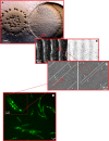
FIG. 3.
Coordinated movements of M. xanthus cells. (A) When M. xanthus cells (left) encounter and then penetrate an E. coli colony (right) they align, forming accordion waves (ripples). In contrast, cells that do not encounter E. coli cells starve and undergo fruiting body formation (dark structures on the left). (The picture of M. xanthus invading an E. coli colony was adapted from reference with permission of Blackwell Publishing Ltd.) (B) Phase-contrast microscopy (left) and fluorescence microscopy (right) of wild-type M. xanthus cells mixed at a 50:1 ratio with GFP-labeled cells, showing that during rippling behavior, the rippling pattern is stably maintained, although individual cells change position. (The picture of the M. xanthus ripple structures was adapted from reference .) (C) Ripples in a monolayer culture. The pictures show that during a collision between two waves, cells in one wave penetrate the opposing wave by one cell length, followed by cell reversals. (The picture of the M. xanthus ripples was adapted from reference .) (D) M. xanthus cells expressing FrzCD-GFP. FrzCD forms clusters that align relative to each other when cells make side-to-side contacts (see inset). We propose that during rippling behavior, side-to-side contacts between cells stimulate the FrzCD receptor to trigger reversals.
FIG. 4.
FIG. 4.
Components of the S-motility system. (A) A model showing the different components of the S-engine. The inner membrane PilC is present at both the leading pole and the lagging pole in equal amounts (19). The ATPase PilB is more abundant at the leading pole, where it catalyzes the polymerization of the TFP by hydrolyzing ATP. PilT is also an ATPase but with an opposing function: it catalyzes the disassembly of the TFP. PilT localizes mostly at the lagging pole. It is also present at the leading cell pole in smaller amounts to power the retraction of the TFP. The combined activities of PilB and PilT lead to the periodic assembly and retraction of TFP, which allow the cell to move forward. The lipoprotein Tgl is present only at the leading pole, and this causes the secretin PilQ to be assembled at the leading pole and disassembled at the lagging pole. The presence of PilQ channels at the leading pole allows the assembly of PilA filaments from the pool of monomers stored in the membrane or synthesized de novo. (B) Polar TFP. (The picture was obtained by atomic force microscopy and adapted from reference .) (C) Dried extracellular matrix material appears as fibrils that interconnect cells when viewed by scanning electron microscopy. (The picture of the M. xanthus fibril material was adapted from reference with permission from Elsevier.)
FIG. 5.
FIG. 5.
Slime secretion model for A motility. A slime polymer (yellow) is synthesized within a putative inner membrane-localized biosynthetic machinery (pink), dehydrated, and introduced into a nozzle (blue) embedded within the peptidoglycan. Water flowing from the extracellular space creates a gradient of hydration leading to the swelling of the hydrogel within the nozzle chamber. Release of the hydrogel at the outer membrane creates a force (black arrow) that pushes the cell forward. (A) EM image of ribbons at a M. xanthus cell pole. (B) Average of side-view projection images of the Phormidium nozzle, a 40-nm-long symmetric open complex with a central hole of variable diameter (8 to 14 nm). (C) Rings observed in the M. xanthus outer membrane. (All panels were adapted from reference with permission from Elsevier.)
FIG. 6.
FIG. 6.
Focal adhesion model for A motility. Focal adhesion complexes (green) are assembled at the leading cell pole along the MreB cytoskeleton (red). The complexes appear to retain fixed positions as an associated motor pulls on the MreB filament. The blow up shows a cartoon of the proposed machinery. An unidirectional engine (green) pulls the MreB filament on one side and is connected to an envelope-spanning protein system (pink) that ends with an adhesin (blue). Movement is produced because the inner and outer semifluidic membranes “flow” through the complex and a machinery-bound peptidoglycan hydrolase (purple) digests the cell wall locally. (A) Gliding motility of a cephalexin-treated cell. The cell unfolds as if it is pulled by its front part while the back part remains inert. The cell is stained with the membrane FM4-64 dye and shows no obvious septa. (The picture of the M. xanthus cephalexin-treated cell was adapted from reference .) (B) Fixed AglZ-YFP clusters in a moving cell. Frames of a movie (30-s intervals) showing a moving M. xanthus cell expressing AglZ-YFP (artificially colored in magenta) are shown. Black arrow, direction of movement. Scale bar = 2 μm. (The picture of the AglZ-YFP-labeled M. xanthus cell was adapted from reference .)
FIG. 7.
FIG. 7.
Schematic diagram of the Mcp7, Dif, Frz, and Che4 chemotaxis systems of M. xanthus. These chemosensory systems are important in the regulation of M. xanthus motility. Chemosensory proteins might form complexes analogously to their enteric counterparts. Dashed arrows indicate the cross talk between the different chemosensory systems.
FIG. 8.
FIG. 8.
The localizations of A- and S-motility proteins change synchronously as cells reverse. Time-lapse fluorescence microscopy of a moving M. xanthus cell expressing FrzS-GFP and RomR-mCherry is shown. The top panels show fluorescence images. The bottom panels show the same cell in bright field. The cartoon shows schematically the asymmetric bipolar localization of FrzS (green) and RomR (red). The FrzS larger cluster is at the cell leading pole while the RomR larger cluster is at the lagging pole. During a reversal, proteins redistribute and appear in equal amount at both poles. As soon as the cell starts moving again, polarity is reestablished and FrzS appears at the new leading pole, whereas RomR localizes at the lagging end of the cell. (The picture of the FrzS-GFP RomR-mCherry-labeled M. xanthus cell was adapted from reference by permission from Macmillan Publishers Ltd.)

Similar articles

Cited by

References

    1. Arnold, J. W., and L. J. Shimkets. 1988. Cell surface properties correlated with cohesion in Myxococcus xanthus. J. Bacteriol. 170:5771-5777. - PMC - PubMed
    1. Astling, D. P., J. Y. Lee, and D. R. Zusman. 2006. Differential effects of chemoreceptor methylation-domain mutations on swarming and development in the social bacterium Myxococcus xanthus. Mol. Microbiol. 59:45-55. - PubMed
    1. Bassler, B. L., and R. Losick. 2006. Bacterially speaking. Cell 125:237-246. - PubMed
    1. Behmlander, R. M., and M. Dworkin. 1994. Biochemical and structural analyses of the extracellular matrix fibrils of Myxococcus xanthus. J. Bacteriol. 176:6295-6303. - PMC - PubMed
    1. Behmlander, R. M., and M. Dworkin. 1994. Integral proteins of the extracellular matrix fibrils of Myxococcus xanthus. J. Bacteriol. 176:6304-6311. - PMC - PubMed

Publication types

MeSH terms

Substances

LinkOut - more resources