Review
. 2010 Jun;74(2):298-340.
doi: 10.1128/MMBR.00005-10.

The evolution of sex: a perspective from the fungal kingdom

Affiliations
Review

The evolution of sex: a perspective from the fungal kingdom

Soo Chan Lee et al. Microbiol Mol Biol Rev. 2010 Jun.

Abstract

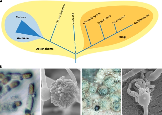
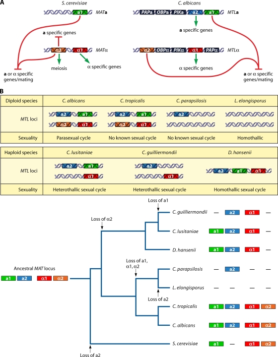
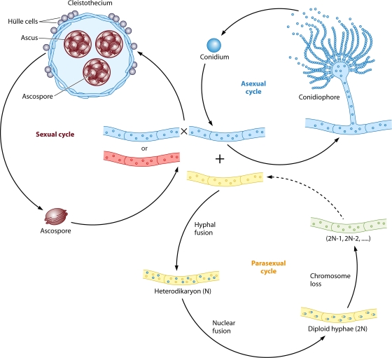
Sex is shrouded in mystery. Not only does it preferentially occur in the dark for both fungi and many animals, but evolutionary biologists continue to debate its benefits given costs in light of its pervasive nature. Experimental studies of the benefits and costs of sexual reproduction with fungi as model systems have begun to provide evidence that the balance between sexual and asexual reproduction shifts in response to selective pressures. Given their unique evolutionary history as opisthokonts, along with metazoans, fungi serve as exceptional models for the evolution of sex and sex-determining regions of the genome (the mating type locus) and for transitions that commonly occur between outcrossing/self-sterile and inbreeding/self-fertile modes of reproduction. We review here the state of the understanding of sex and its evolution in the fungal kingdom and also areas where the field has contributed and will continue to contribute to illuminating general principles and paradigms of sexual reproduction.


Figures

FIG. 1.
FIG. 1.
Phylogeny of fungi and metazoans in the eukaryotic opisthokonts and sexual structures of four major fungal phyla. (A) The animal and fungal kingdoms are derived from a common ancestor, forming a clade called the opisthokonts. The opisthokonts contain several unicellular lineages (i.e., choanoflagellates and nuclearia). Within the four fungal phyla, the chytridiomycetes and zygomycetes form the basal lineages, and the ascomycetes and basidiomycetes are more recently diverged as the monophyletic dikarya. (B) The four major phyla have their own characteristics in sexual development. From left to right are sexual hyphae of the chytridiomycete Allomyces macrogynus, in which the male is orange and the female is hyaline, from which flagellated gametes arise; a zygospore of Mucor circinelloides, a pathogenic zygomycete; cleistothecia during sexual development in the ascomycete Aspergillus nidulans, which harbor sexual spores; and basidia and sexual spore chains of the human-pathogenic basidiomycete Cryptococcus neoformans.
FIG. 2.
FIG. 2.
MAT and MTL loci of S. cerevisiae and C. albicans and the evolutionary trajectory of the MTL locus and sexuality within the Candida clade. (A) The MAT locus of S. cerevisiae and the MTL locus of C. albicans share similar architectures. Compared to S. cerevisiae, C. albicans has additional genes, a2, PAP, OBP, and PIK, in the MTL locus. (B) Compositions of the MTL loci of diploid and haploid Candida spp. vary. During speciation within the Candida clade, multiple independent losses of the ancestral MAT locus components a1, a2, α1, and α2 and transitions in sexuality have occurred (see also reference 274). The additional genes are not presented. The tree was redrawn based on a six-gene phylogeny (50).
FIG. 3.
FIG. 3.
Life cycle of A. nidulans. A. nidulans can undergo three life cycles. First, it produces conidia robustly during an asexual cycle. A conidium germinates to form hyphae, from which conidiophores develop to produce more conidia. Second, it can undergo a homothallic sexual cycle involving selfing or out-crossing to generate fruiting bodies (called cleistothecia) containing thousands of ascospores, which germinate to form hyphae. Third, it can undergo a parasexual cycle. Heterohyphae fuse to form a heterodikaryon, followed by nuclear fusion to generate diploid hyphae, in which random chromosome loss occurs to restore the haploid chromosome number.
FIG. 4.
FIG. 4.
Sexual structures of A. fumigatus. Shown are scanning electron micrographs of cleistothecia (A), an ascus with eight ascospores (B), and ascospores (C). Scale bars represent 100 μm (A), 2 μm (B), and 2 μm (C). (Courtesy of Celine O'Gorman.)
FIG. 5.
FIG. 5.
Model of evolution of MAT loci within aspergilli. (A) In the model of homothallism as ancestral, the last common ancestor contained both alpha box and HMG domain genes adjacent to each other flanked by the SLA2 and APN1 genes. Next, in one lineage, a chromosomal break and translocation occurred to rearrange the alpha box and HMG domain genes to different chromosomes flanked by either the SLA2 or APN1 gene, giving rise to extant homothallic species. In addition, in an alternative lineage, chromosome segregation and gene loss occurred to maintain either the alpha box or HMG domain genes at the original locus, giving rise to extant heterothallic species. (B) In the model of heterothallism as ancestral, the last common ancestor contained either the alpha box or the HMG domain gene at the same locus flanked by the SLA2 and APN1 genes. This species underwent a chromosomal break and translocation to rearrange the alpha box and HMG genes to different chromosomes flanked by the SLA2 and APN1 genes, respectively, to evolve into extant homothallic species (A. nidulans). In addition, this ancestor also underwent gene duplication and chromosomal translocation to maintain either the alpha box or HMG domain gene at both the original locus and an unlinked locus with modified flanking genes to evolve into other homothallic species (N. fischeri).
FIG. 6.
FIG. 6.
MAT loci of the dermatophytes and dimorphic fungi. The phylogenetic organization of dermatophytes and dimorphic fungi was deduced from partial 18S rRNA gene and internal transcribed spacer (ITS) sequences. The MAT locus of Coccidioides species expanded (broken red line) by the capture of the APN2 and COX13 genes into the MAT locus, which typically flank the MAT locus in other fungal species. This analysis also revealed a unique gene arrangement of the MAT locus and flanking region of the dermatophytes in which the APN2 and COX13 genes lie on the same side as the SLA2 gene. P. marneffei also has a small MAT locus (3.3 kb) similar in size to that of the dermatophytes and much smaller than those of the dimorphic fungi. In B. dermatitidis, the SLA2 gene is located more than 50 kb away from the HMG domain gene in the MAT1-2 locus, but the size of MAT is as yet unknown, as the MAT1-1 idiomorph has not yet been defined. T. equinum, Trichophyton equinum; T. tonsurans, Trichophyton tonsurans; T. rubrum, Trichophyton rubrum; M. canis, Microsporum canis.
FIG. 7.
FIG. 7.
MAT loci of the tetrapolar basidiomycete U. maydis and bipolar basidiomycetes U. hordei, M. globosa, and C. neoformans. (A) The pheromone/pheromone receptor and transcription factor loci are unlinked in U. maydis. There are two known a loci, containing the mfa and pra genes. Two representative b loci (of an estimated ∼25 loci) are presented, which encode two divergently transcribed homeodomain proteins (HD1 and HD2). (B) However, the pheromone/pheromone receptor and transcription factor genes are linked in the U. hordei, M. globosa, and C. neoformans genomes. Many additional genes have been incorporated into the C. neoformans MAT locus. (The MAT locus alleles of the serotype D strains JEC21 for the α mating type and JEC20 for the a mating type are presented.)
FIG. 8.
FIG. 8.
Model of transition to extant bipolar systems from an ancestral tetrapolar system in basidiomycete MAT loci. Linkage of the a and b loci generates an extended MAT locus in the U. hordei and M. globosa genomes. In the C. neoformans genome, gene acquisition occurred into the MAT locus, and the two loci were then linked, generating an intermediate tripolar system in which the a and b loci were linked in one mating type MAT allele with the other mating alleles unlinked, followed by gene conversion and chromosome rearrangement to generate the extant extended bipolar MAT locus.
FIG. 9.
FIG. 9.
Sex recognition systems and evolutionary trajectory in C. neoformans sibling species. The clade of pathogenic Cryptococcus species is characterized as having bipolar mating systems. Only one of the genes for HD1 or HD2 transcription factors is found in the a and α MAT alleles. However, the C. amylolentus, T. wingfieldii, and C. heveanensis MAT loci contain both HD1 and HD2 genes, suggesting that two paired, divergently oriented HD1 and HD2 genes in the MAT locus are the ancestral arrangement. Interestingly, F. depauperata grows only as a filamentous form resembling the Cryptococcus sexual state, indicating that this fungus may be an obligate sexual species. Fungi in the Tremellales group retain the tetrapolar mating system. One species in the Kwoniella clade is sexual and bipolar or tetrapolar. Thus, bipolarity may have emerged during the origin of the Kwoniella and Filobasidella clades from a common tetrapolar ancestor. The tree was drawn based on six-gene MLST as described previously by Findley et al. (57). C. dejecticola, Cryptococcus dejecticola; B. dendrophila, Bullera dendrophila; K. mangroviensis, Kwoniella mangroviensis. C. bestiolae, Cryptococcus bestiolae; T. globispora, Tremella globispora; C. humicola, Cryptococcus humicola.
FIG. 10.
FIG. 10.
Pathway of trisporic acid synthesis in zygomycetes. In the (+) strains, 4-dihydromethyl is produced from β-carotene, secreted, and taken up by the (−) strains, where it is finally converted to trisporic acid. Trisporin and trisporol are produced in the (−) strains, exported, and then imported by the (+) strains and converted to trisporic acid.
FIG. 11.
FIG. 11.
sex loci in the genomes of three zygomycetes. The sex loci in the three zygomycetes M. circinelloides, P. blakesleeanus, and R. oryzae are within a gene cluster encompassing the TPT, HMG, and RNA helicase genes. The sex loci encode idiomorphic HMG transcription factors, SexP and SexM, for (+) and (−) mating types, respectively. The P. blakesleeanus (+) sex locus contains a repetitive element, and the R. oryzae (+) sex locus carries an additional ORF in the gene cluster. The orientation of the sex genes is divergent in P. blakesleeanus and the TPT genes in the R. oryzae sex locus alleles are inverted compared to those of M. circinelloides and P. blakesleeanus. Gray boxes indicate the extent of the sex locus. Note that the promoter of the TPT gene in M. circinelloides is within the border of the sex locus. BTB, bric-a-brac, tramtrack, and broad complex.
FIG. 12.
FIG. 12.
Models for the mode of action of SexP and SexM. First, SexP and SexM may function in distinct promoter areas with different regulatory complexes to converge on common target genes (model I). Second, SexP and SexM may interact to form a heterodimer to function to transcribe mating genes (model II). Third, SexP and SexM may regulate different subsets of mating genes (model III). Note that SexP and SexM likely function both pre- and postfusion.
FIG. 13.
FIG. 13.
Early steps in sex chromosome evolution. The P. blakesleeanus sex locus supports the hypothesis posited by Ohno (222): a sex determinant gene arises on an autosome and undergoes gene inversion followed by gene divergence in the two alleles (top). A repetitive element is inserted at the locus and elsewhere on the proto-sex chromosome. The sex locus expands to form an early sex chromosome. However, the sex loci in M. circinelloides and R. oryzae present an alternative scenario in which sex genes in the same orientation could be an ancestral character, and therefore, gene inversion might follow rather than precede gene divergence (bottom).
FIG. 14.
FIG. 14.
The microsporidian sex-related locus. (A) Differential interference contrast (DIC) and transmission electron microscopy (TEM) images of an E. cuniculi spore. A polar tube is everted from the spore. The spore contains a coiled polar tube. (B) sex-related loci observed for four different microsporidian species. The E. cuniculi and A. locustae genomes contain a syntenic region encompassing TPT (green), HMG (red), and RNA helicase (blue) genes similar to that observed at the zygomycete sex loci (Fig. 11). The E. cuniculi sex-related locus has an additional weak homology HMG gene (purple). All sex-related loci from the four different microsporidian species have a conserved hypothetical protein gene (yellow). Two other microsporidians, E. bieneusi and N. ceranae, have an unlinked RNA helicase gene. Only microsporidia and zygomycetes share the synteny of the TPT, HMG, and RNA helicase genes compared to other fungi for which complete genome sequences are available.
FIG. 15.
FIG. 15.
Evolutionary trajectory of the sex locus within zygomycetes and microsporidia. The ancestral sex loci are hypothesized to have contained diverged alleles of the TPT, HMG, and RNA helicase genes. By gene eviction from the sex locus, only the TPT or the RNA helicase gene was retained flanking the sex locus. In this evolutionary model, the microsporidian TPT and RNA helicase genes are paralogs of those that flank the zygomycete sex locus.
FIG. 16.
FIG. 16.
The Allomyces macrogynus life cycle. The sporophyte generation is maintained asexually through the production of diploid zoospores. Meiosporangia that develop on the mature sporophyte produce meiospores, haploid uniflagellate spores that produce mature gametophytes capable of forming male and female gametangia. Gametes are released and undergo cell fusion (plasmogamy), followed by nuclear fusion (karyogamy), to form a biflagellate uninucleate zygote that develops into a sporophytic plant.
FIG. 17.
FIG. 17.
Chytrids produce a novel mating pheromone. (A) Chemical structure of sirenin. (B) Bioassay design for sirenin chemotaxis. Female gametangia are placed into the center well of a thin agar plate. Experimental groups are placed into outlying wells. If chemotaxis toward sirenin occurs, clusters toward the center well are observed. The orange circles represent male gametangia, white circles represent female gametangia, and gray circles represent zoosporangia.
FIG. 18.
FIG. 18.
Illustration of the same-sex mating process in the two human-pathogenic fungi C. neoformans and C. albicans. Same-sex mating in C. neoformans occurs between two α cells, whereas conventional mating involves two opposite-mating-type cells. C. albicans a/a diploid cells also need to be opaque to undergo same-sex mating. In the absence of Bar1 protease activity or in the presence of α cells, the a/a diploids undergo homothallic mating. The 4N cells then undergo a parasexual cycle to return to a diploid state as seen in opposite-sex mating.
FIG. 19.
FIG. 19.
Evolution of transcription factors encoded by the fungal MAT locus. Zygomycetes, representative basal fungi, utilize an HMG transcription factor for sexual recognition, which parallels the human sex determination system with the Sry HMG protein. The common ancestor to the opisthokont clade may have had an HMG protein as a sex determinant (52). Alternatively, given the ubiquity of both HMG and homeodomain (HD) factors, there may have been two ancestral sex-determining systems that competed for preeminence, or in some cases, such as C. albicans, both remain resident at the MAT locus. There were multiple losses of HMG as a sex determinant, and in some lineages, α domain and/or HD transcription factors were adopted. Basidiomycetes lost the MAT-encoded HMG but acquired HD factors as sex determinants. In addition, they adapted multiallelism (e.g., U. maydis and C. cinerea). However, some basidiomycetes have returned to the biallelic bipolar state (e.g., C. neoformans, U. hordei, and M. globosa). The α domain proteins were likely incorporated into MAT during the evolution of the ascomycete MAT loci but also share sequence identity with HMG domains and may therefore be distantly related. In Saccharomyces spp., the HMG protein no longer functions as a sex determinant, and the mating type switching system evoked by the Ho endonuclease was recently acquired within this group. In contrast, several species of the Candida CTG clade retained the HMG gene in the MTL locus, and thus, three classes of transcription factors are present (HMG, HD, and alpha box), and no silent cassette or mating type switching occurs.

Similar articles

Cited by

References

    1. Adams, T. H., J. K. Wieser, and J. H. Yu. 1998. Asexual sporulation in Aspergillus nidulans. Microbiol. Mol. Biol. Rev. 62:35-54. - PMC - PubMed
    1. Alby, K., D. Schaefer, and R. J. Bennett. 2009. Homothallic and heterothallic mating in the opportunistic pathogen Candida albicans. Nature 460:890-893. - PMC - PubMed
    1. Alexopoulos, C. J. 1962. Introductory mycology, 2nd ed. John Wiley & Sons, New York, NY.
    1. Alexopoulos, C. J., C. W. Mims, and M. Blackwell. 1996. Introductory mycology, 4th ed. John Wiley & Sons, New York, NY.
    1. Alvarez-Perez, S., J. L. Blanco, P. Alba, and M. E. Garcia. 2009. Mating type and invasiveness are significantly associated in Aspergillus fumigatus. Med. Mycol. 26:1-6. - PubMed

Publication types