Review
. 2010 Jun;22(6):413-9.
doi: 10.1093/intimm/dxq047.

Finding the right niche: B-cell migration in the early phases of T-dependent antibody responses

Affiliations
Review

Finding the right niche: B-cell migration in the early phases of T-dependent antibody responses

João Pedro Pereira et al. Int Immunol. 2010 Jun.

Abstract

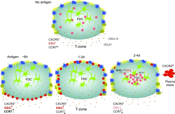
Humoral immune responses depend on B cells encountering antigen, interacting with helper T cells, proliferating and differentiating into low-affinity plasma cells or, after organizing into a germinal center (GC), high-affinity plasma cells and memory B cells. Remarkably, each of these events occurs in association with distinct stromal cells in separate subcompartments of the lymphoid tissue. B cells must migrate from niche to niche in a rapid and highly regulated manner to successfully mount a response. The chemokine, CXCL13, plays a central role in guiding B cells to follicles whereas T-zone chemokines guide activated B cells to the T zone. Sphingosine-1-phosphate (S1P) promotes cell egress from the tissue, as well as marginal-zone B-cell positioning in the spleen. Recent studies have identified a role for the orphan receptor, EBV-induced molecule 2 (EBI2; GPR183), in guiding activated B cells to inter and outer follicular niche(s) and down-regulation of this receptor is essential for organizing cells into GCs. In this review, we discuss current understanding of the roles played by chemokines, S1P and EBI2 in the migration events that underlie humoral immune responses.


Figures

Fig. 1.
Fig. 1.
Positioning of activated antigen-specific B cells in inter and outer follicular niches of spleen and lymph node (LN) at days 2–3 of the response. Five to 10 million MD4 Ig-transgenic B cells that express IgMa and IgDa specific for hen egg lysozyme (HEL), and ovalbumin (OVA)-specific OTII TCR transgenic T cells were co-transferred into C57BL/6 mice immunized with 50 μg of HEL–OVA in Ribi adjuvant (Sigma) intraperitoneally for 2 days (spleen) or subcutaneously for 3 days (LN). Cryostat sections were stained with antibodies to B220 or IgD to detect all follicular B cells (brown) and to IgDa or IgMa + IgDa to detect the activated MD4 B cells (blue). Outer, Center and Inter refer to regions of the follicle. Scale bar is 100 μm.
Fig. 2.
Fig. 2.
Model of EBI2 ligand distribution and B-cell migration in follicles during the early phase of T-cell-dependent antibody responses in spleen and lymph nodes. Naive B cells (pink circles) express CXCR5 and EBI2, reside in follicles and migrate over the processes of FDCs (green), MRCs (yellow), CD169+ macrophages (dark blue) and likely other stromal cells (not depicted). FDCs and MRCs produce CXCL13 (small green dots) and EBI2 ligand is suggested to be made by cells at the follicle perimeter, particularly in inter and outer follicular regions (blue–green gradient, where dark shading represents areas of highest ligand concentration). B-cell movement within the FDC-rich area is largely dependent on CXCR5 and CXCL13 and is critical for antigen scanning. Within 6 h after encountering cognate antigen, activated B cells (red circles) up-regulate CCR7 and EBI2. Activated B cells become more responsive to CCL21 (orange dots) and most likely to EBI2 ligand, while maintaining CXCR5 responsiveness. Balanced chemoattraction mediated by CXCR5 and CCR7, and possibly also responsiveness to local EBI2 ligand, promotes uniform distribution of activated B cells along the interface between follicles and T-cell areas (B/T zone), where cognate interactions with antigen-specific T cells are initiated. Between 1 and 2 days later, B cells down-regulate CCR7 while maintaining high levels of EBI2 (and CXCR5) and move to inter and outer follicular regions where EBI2 ligand concentration might be highest (dark shading). Inter and outer follicular niches are characterized by layers of CD169+ macrophages and MRCs and the interfollicular niche also contains dendritic cells (brown cells). B-cell proliferation and commitment to plasmablast or GC differentiation pathway ensues, perhaps influenced by signals received from local niche cells. Some activated B cells differentiate into plasmablasts and accumulate at extrafollicular sites in a CXCR4-dependent manner. Other activated B cells up-regulate Bcl6, which represses Ebi2, and take on a GC fate. GC-committed B cells move to the center follicle due to loss of EBI2 and perhaps attracted by CXCL13 or another FDC-derived cue and initiate the GC reaction that will produce high-affinity memory B cells and plasma cells.

References

    1. Cyster JG, Ansel KM, Reif K, et al. Follicular stromal cells and lymphocyte homing to follicles. Immunol. Rev. 2000;176:181. - PubMed
    1. Allen CD, Cyster JG. Follicular dendritic cell networks of primary follicles and germinal centers: phenotype and function. Semin. Immunol. 2008;20:14. - PMC - PubMed
    1. Tew JG, Wu J, Qin D, Helm S, Burton GF, Szakal AK. Follicular dendritic cells and presentation of antigen and costimulatory signals to B cells. Immunol. Rev. 1997;156:39. - PubMed
    1. Katakai T, Suto H, Sugai M, et al. Organizer-like reticular stromal cell layer common to adult secondary lymphoid organs. J. Immunol. 2008;181:6189. - PubMed
    1. Mueller SN, Germain RN. Stromal cell contributions to the homeostasis and functionality of the immune system. Nat. Rev. Immunol. 2009;9:618. - PMC - PubMed

Publication types

Substances