. 2010 Jul;11(7):555-60.
doi: 10.1038/embor.2010.79. Epub 2010 May 28.

Nucleosome occupancy landscape and dynamics at mouse recombination hotspots

Affiliations

Nucleosome occupancy landscape and dynamics at mouse recombination hotspots

Irina V Getun et al. EMBO Rep. 2010 Jul.

Abstract

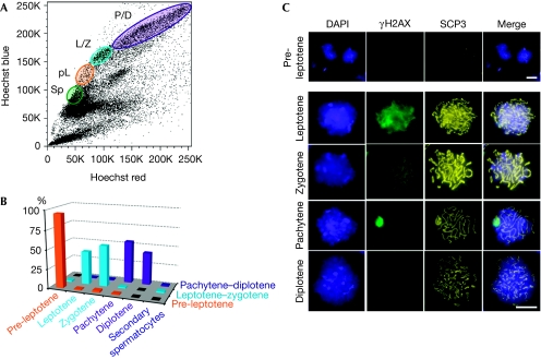
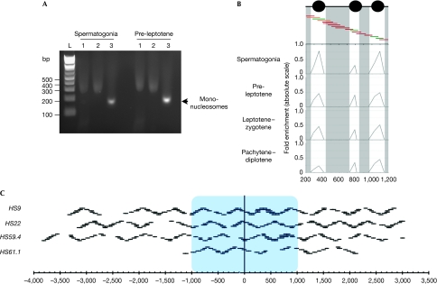
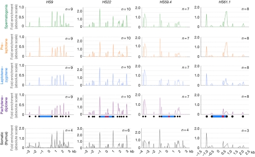
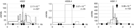
During meiosis, paternal and maternal homologous chromosomes recombine at specific recombination sites named hotspots. What renders 2% of the mammalian genomes permissive to meiotic recombination by allowing Spo11 endonuclease to initiate double-strand breaks is largely unknown. Work in yeast has shown that chromatin accessibility seems to be important for this activity. Here, we define nucleosome profiles and dynamics at four mouse recombination hotspots by purifying highly enriched fractions of meiotic cells. We found that nucleosome occupancy is generally stable during meiosis progression. Interestingly, the cores of recombination hotspots have largely open chromatin structure, and the localization of the few nucleosomes present in these cores correlates precisely with the crossover-free zones in recombinogenic domains. Collectively, these high-resolution studies suggest that nucleosome occupancy seems to direct, at least in part, how meiotic recombination events are processed.


Conflict of interest statement

The authors declare that they have no conflict of interest.

Figures

Figure 1
Figure 1
Fluorescence-activated cell sorting purification and characterization of meiotic-stage cells from adult mice. (A) A representative Hoechst FACS profile, with the meiotic cell populations indicated. For this study, the four populations purified were spermatogonia (Sp), pre-leptotene (pL), leptotene–zygotene (L/Z) and pachytene–diplotene (P/D). (B) Graph indicating the percentage of each cell type per purified population: pre-leptotene (95%), leptotene–zygotene (43–53%) and pachytene–diplotene (42–55%). (C) Representative pictograms of meiotic cells of the purified fractions. Cells were counterstained with DAPI and co-immunostained with γH2AX and SCP3 to confirm the stage of the FACS-sorted meiotic cell population. γH2AX shows the diffuse staining before pachytene, where it becomes highly enriched on the XY-sex body. SCP3 becomes clearly visible at leptotene and zygotene, and at pachytene it forms perfectly along the length of the homologous chromosomes, but then it begins to disintegrate at diplotene. Scale bars, 20 μm. DAPI, 4,6-diamidino-2-phenylindole; γH2AX, phosphorylated histone H2AX; FACS, fluorescence-activated cell sorting; SCP3, synaptonemal complex protein 3.
Figure 2
Figure 2
Schematic of the nucleosome mapping technique. (A) Typical mono-nucleosome preparation from the indicated FACS-purified meiotic cells is shown. Lanes 1 correspond to the total chromatin fraction after MNAse digestion, lanes 2 indicate the extracted nucleosomes after native chromatin treatment and lanes 3 show the purified mono-nucleosomes after desalting. The high salt concentration causes aberrant migration in lanes 1 and 2. (B) FACS-purified meiotic fractions are digested to completion with MNase, DNA is purified and mono-nucleosome fragments are isolated. Mono-nucleosomal DNA is used as a template for quantitative PCR with overlapping primer pairs. A small 1 kb region is shown at the HS9 locus. Protection of DNA from MNase digestion by nucleosomes will result in a robust PCR signal (green primer pairs), partial protection in an intermediate PCR signal (orange) and no protection in a trace PCR signal (dark red). Profiles are shown at the four studied stages as indicated. (C) Diagram representing all the primer pairs used for the four studied hotspots. The blue box indicates the approximate recombinogenic core. Gaps in the oligonucleotide pairs were owing to simple repeats, poor efficiency or primer pair failure to yield unique products. FACS, fluorescence-activated cell sorting; MNase, micrococcal nuclease.
Figure 3
Figure 3
Nucleosome occupancy at the four meiotic hotspots during meiotic progression. Nucleosome profiles at the recombination hotpots in spermatogonia, pre-leptotene, leptotene–zygotene and pachytene–diplotene meiotic fractions, as well as in somatic primary thymic cells, are shown for each of the indicated hotspots. Profiles are absolute, with the vertical scale being the fold enrichment between the mono-nucleosome MNase-digested DNA fraction and undigested genomic DNA, which is calculated by using the formula 2−ΔCt. The x-axis represents the location across the analysed locus. Nucleosome positions outside (black) or within (red) hotspot cores (blue) are indicated. The numbers of independent experimental repeats for each profile are shown. MNase, micrococcal nuclease.
Figure 4
Figure 4
Nucleosome positioning within the hotspot core is correlated with the crossover repulsion zones. Recombination profiles of HS22, HS59.4 and HS61.1 are shown. Both CO orientations are shown with B6-to-DBA (black) and DBA-to-B6 (grey); only the latter orientation is shown for HS59.4. Recombination rates and the number of molecules analysed to obtain the CO profiles at each hotspot are indicated. Nucleosomes are drawn to scale. Black arrows indicate the CO refractory zones within the hotspot cores (Wu et al, 2010). CO, crossover.

References

    1. Bastos H, Lassalle B, Chicheportiche A, Riou L, Testart J, Allemand I, Fouchet P (2005) Flow cytometric characterization of viable meiotic and postmeiotic cells by Hoechst 33342 in mouse spermatogenesis. Cytometry A 65: 40–49 - PubMed
    1. Bois PR (2007) A highly polymorphic meiotic recombination mouse hotspot exhibits incomplete repair. Mol Cell Biol 27: 7053–7062 - PMC - PubMed
    1. Borde V, Robine N, Lin W, Bonfils S, Geli V, Nicolas A (2009) Histone H3 lysine 4 trimethylation marks meiotic recombination initiation sites. EMBO J 28: 99–111 - PMC - PubMed
    1. Buard J, Barthes P, Grey C, de Massy B (2009) Distinct histone modifications define initiation and repair of meiotic recombination in the mouse. EMBO J 28: 2616–2624 - PMC - PubMed
    1. Di Felice F, Chiani F, Camilloni G (2008) Nucleosomes represent a physical barrier for cleavage activity of DNA topoisomerase I in vivo. Biochem J 409: 651–656 - PubMed

Publication types

LinkOut - more resources