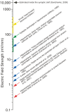
Field effects in the CNS play functional roles
- PMID: 20508749
- PMCID: PMC2876880
- DOI: 10.3389/fncir.2010.00015
Field effects in the CNS play functional roles
Abstract
An endogenous electrical field effect, i.e., ephaptic transmission, occurs when an electric field associated with activity occurring in one neuron polarizes the membrane of another neuron. It is well established that field effects occur during pathological conditions, such as epilepsy, but less clear if they play a functional role in the healthy brain. Here, we describe the principles of field effect interactions, discuss identified field effects in diverse brain structures from the teleost Mauthner cell to the mammalian cortex, and speculate on the function of these interactions. Recent evidence supports that relatively weak endogenous and exogenous field effects in laminar structures reach significance because they are amplified by network interactions. Such interactions may be important in rhythmogenesis for the cortical slow wave and hippocampal sharp wave-ripple, and also during transcranial stimulation.
Keywords: LFP; computation; ephaptic; field effect; neurotransmission; transcranial stimulation.
Figures


Similar articles
-
Coordinated Interaction between Hippocampal Sharp-Wave Ripples and Anterior Cingulate Unit Activity.J Neurosci. 2016 Oct 12;36(41):10663-10672. doi: 10.1523/JNEUROSCI.1042-16.2016. J Neurosci. 2016. PMID: 27733616 Free PMC article.
-
Slow periodic activity in the longitudinal hippocampal slice can self-propagate non-synaptically by a mechanism consistent with ephaptic coupling.J Physiol. 2019 Jan;597(1):249-269. doi: 10.1113/JP276904. Epub 2018 Nov 10. J Physiol. 2019. PMID: 30295923 Free PMC article.
-
Ephaptic Coupling of Cortical Neurons: Possible Contribution of Astroglial Magnetic Fields?Neuroscience. 2018 Feb 1;370:37-45. doi: 10.1016/j.neuroscience.2017.07.072. Epub 2017 Aug 6. Neuroscience. 2018. PMID: 28793233 Review.
-
Stimulating forebrain communications: Slow sinusoidal electric fields over frontal cortices dynamically modulate hippocampal activity and cortico-hippocampal interplay during slow-wave states.Neuroimage. 2016 Jun;133:189-206. doi: 10.1016/j.neuroimage.2016.02.070. Epub 2016 Mar 3. Neuroimage. 2016. PMID: 26947518
-
Endogenous and exogenous electric fields as modifiers of brain activity: rational design of noninvasive brain stimulation with transcranial alternating current stimulation.Dialogues Clin Neurosci. 2014 Mar;16(1):93-102. doi: 10.31887/DCNS.2014.16.1/ffroehlich. Dialogues Clin Neurosci. 2014. PMID: 24733974 Free PMC article. Review.
Cited by
-
Universal theory of brain waves: from linear loops to nonlinear synchronized spiking and collective brain rhythms.Phys Rev Res. 2020 Apr-Jun;2(2):023061. doi: 10.1103/PhysRevResearch.2.023061. Epub 2020 Apr 21. Phys Rev Res. 2020. PMID: 33718881 Free PMC article.
-
Contribution of transcranial oscillatory stimulation to research on neural networks: an emphasis on hippocampo-neocortical rhythms.Front Hum Neurosci. 2013 Sep 26;7:614. doi: 10.3389/fnhum.2013.00614. Front Hum Neurosci. 2013. PMID: 24133431 Free PMC article. Review.
-
High Frequency Oscillations in Rat Hippocampal Slices: Origin, Frequency Characteristics, and Spread.Front Neurol. 2020 Apr 22;11:326. doi: 10.3389/fneur.2020.00326. eCollection 2020. Front Neurol. 2020. PMID: 32390935 Free PMC article.
-
Computational study of synchrony in fields and microclusters of ephaptically coupled neurons.J Neurophysiol. 2015 May 1;113(9):3229-41. doi: 10.1152/jn.00546.2014. Epub 2015 Feb 11. J Neurophysiol. 2015. PMID: 25673735 Free PMC article.
-
Controlling the local extracellular electric field can suppress the generation and propagation of seizures and spikes in the hippocampus.Brain Stimul. 2025 Mar-Apr;18(2):225-234. doi: 10.1016/j.brs.2025.02.001. Epub 2025 Feb 10. Brain Stimul. 2025. PMID: 39938862 Free PMC article.
References
-
- Arvanitaki A. (1942). Effects evoked in an axon by the activity of a contiguous one. J. Neurophysiol. 5, 81–108
-
- Autere A. M., Lamsa K., Kaila K., Taira T. (1999). Synaptic activation of GABAA receptors induces neuronal uptake of Ca2+ in adult rat hippocampal slices. J. Neurophysiol. 81, 811–816 - PubMed
LinkOut - more resources
Full Text Sources
Other Literature Sources

