. 2010:2010:686457.
doi: 10.1155/2010/686457. Epub 2010 May 24.

Skeletal dysplasias associated with mild myopathy-a clinical and molecular review

Affiliations

Skeletal dysplasias associated with mild myopathy-a clinical and molecular review

Katarzyna A Piróg et al. J Biomed Biotechnol. 2010.

Abstract

Musculoskeletal system is a complex assembly of tissues which acts as scaffold for the body and enables locomotion. It is often overlooked that different components of this system may biomechanically interact and affect each other. Skeletal dysplasias are diseases predominantly affecting the development of the osseous skeleton. However, in some cases skeletal dysplasia patients are referred to neuromuscular clinics prior to the correct skeletal diagnosis. The muscular complications seen in these cases are usually mild and may stem directly from the muscle defect and/or from the altered interactions between the individual components of the musculoskeletal system. A correct early diagnosis may enable better management of the patients and a better quality of life. This paper attempts to summarise the different components of the musculoskeletal system which are affected in skeletal dysplasias and lists several interesting examples of such diseases in order to enable better understanding of the complexity of human musculoskeletal system.


Figures
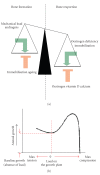
Figure 1
Figure 1
(a) A schematic representation of processes that may influence bone formation and resorption [3]. (b) A graph illustrating the Hueter-Volkman law [4].
Figure 2
Figure 2
A histological H&E (haematoxylin and eosin) stained image of an adult mouse growth plate and a schematic representation of differentiation zones in the tissue. In the growth plate several distinct structural zones can be identified, reflecting the gradual transition of cells through different stages of differentiation [13]. Resting zone acts as a reserve of precursor cells for the proliferating chondrocytes in the columns [10]. Proliferating zone is where the cells flatten and divide, laying down a cartilage extracellular matrix that will later serve as a scaffold for bone formation [14]. In the prehypertrophic zone, the cells enter the maturation zone and begin to enlarge. In the hypertrophic zone, the chondrocytes and their lacunae become 5–12 times bigger [14]. These cells eventually die, triggering vascularisation and bone formation.
Figure 3
Figure 3
(a) A schematic representation of tendon hierarchical structure and a transmission electron microscopy image of a tenocyte embedded in the collagen matrix. (b) A histological (Gomori trichrome, staining collagenous tissues blue) image of a longitudinal section of patellar tendon showing parallel running collagen bundles in the tissue.
Figure 4
Figure 4
Schematic representation of the skeletal muscle structure and histological images of a longitudinal section of murine skeletal muscle and of murine Achilles tendon myotendinous junction (MTJ) stained with a trichrome Gomori stain to visualise collagenous tissues (staining muscle red, collagenous tissues blue, and nuclei black).
Figure 5
Figure 5
Grip strength measurement in COMP-CTD T585M knock-in mice and COMP-T3 ΔD469 knock-in mice at 3 weeks of age. COMP-CTD mice were getting tired and let go of the apparatus easier than their wild type controls, although they were not generally weaker at 3 weeks of age, as seen by the maximum strengths registered (n = 15) [71]. COMP D469 mice were not getting tired and had the same maximum strength as the wild type controls at 3 weeks of age; however, by 9 weeks they were significantly weaker than their wild type littermates and tired easier, which is indicative of a mild myopathy (n = 5). Key: wt: wild type, mut: homozygous for the mutation, *P < .05, **P < .01, ***P < .001 (independent samples t-test).

References

    1. Francomano CA, McIntosh I, Wilkin DJ. Bone dysplasias in man: molecular insights. Current Opinion in Genetics and Development. 1996;6(3):301–308. - PubMed
    1. Mogayzel PJ, Marcus CL. Skeletal dysplasias and their effect on the respiratory system. Paediatric Respiratory Reviews. 2001;2(4):365–371. - PubMed
    1. Harada S-I, Rodan GA. Control of osteoblast function and regulation of bone mass. Nature. 2003;423(6937):349–355. - PubMed
    1. Frost HM. Biomechanical control of knee alignment: some insights from a new paradigm. Clinical Orthopaedics and Related Research. 1997;(335):335–342. - PubMed
    1. Royce PM, Heinmann B. Connective Tissue and Its Heritable Disorders. 2nd edition. Zurich, Switzerland: John Wiley & Sons; 2002.

LinkOut - more resources