Review
. 2010:2010:513948.
doi: 10.1155/2010/513948. Epub 2010 May 23.

Release of inflammatory mediators by human adipose tissue is enhanced in obesity and primarily by the nonfat cells: a review

Affiliations
Review

Release of inflammatory mediators by human adipose tissue is enhanced in obesity and primarily by the nonfat cells: a review

John N Fain. Mediators Inflamm. 2010.

Abstract

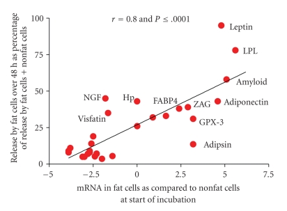
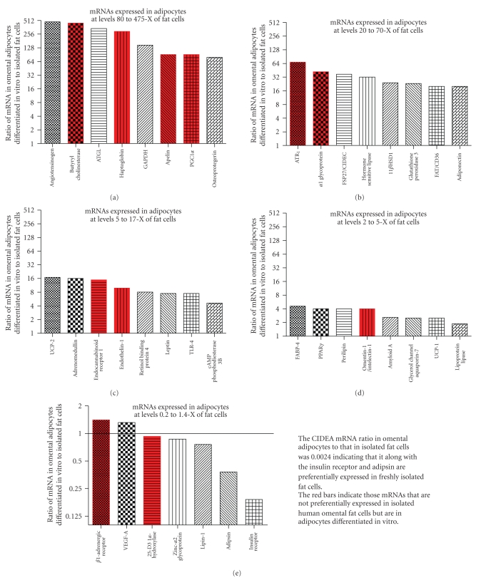
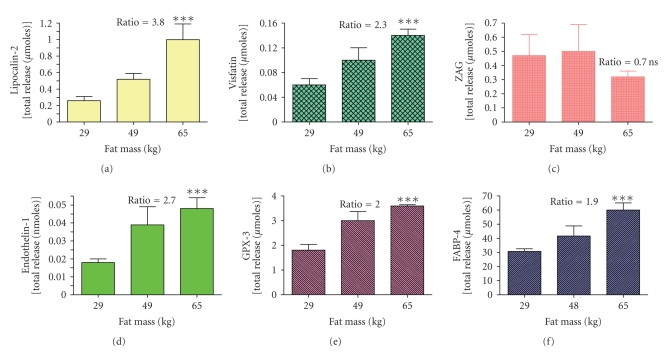
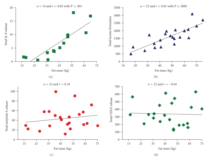
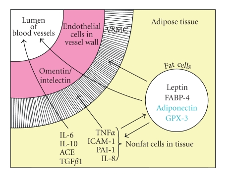
This paper considers the role of putative adipokines that might be involved in the enhanced inflammatory response of human adipose tissue seen in obesity. Inflammatory adipokines [IL-6, IL-10, ACE, TGFbeta1, TNFalpha, IL-1beta, PAI-1, and IL-8] plus one anti-inflammatory [IL-10] adipokine were identified whose circulating levels as well as in vitro release by fat are enhanced in obesity and are primarily released by the nonfat cells of human adipose tissue. In contrast, the circulating levels of leptin and FABP-4 are also enhanced in obesity and they are primarily released by fat cells of human adipose tissue. The relative expression of adipokines and other proteins in human omental as compared to subcutaneous adipose tissue as well as their expression in the nonfat as compared to the fat cells of human omental adipose tissue is also reviewed. The conclusion is that the release of many inflammatory adipokines by adipose tissue is enhanced in obese humans.


Figures

Figure 1
Figure 1
The correlation between releases of 30 adipokines over 48 hours incubation by fat cells isolated from human adipose tissue as compared to gene expression of these adipokines at the start of the incubation. The release data are from Table 1 and expressed as release by fat cells as % of that by fat cells plus nonfat cells over 48 hours. The data for mRNA are derived from those shown in Table 2 except that they are plotted as the ΔCp for the difference between mRNA in fat cells and nonfat cells instead of the ratios, which are derived from the ΔCp values. Data are not included for resistin, CRP and IL-18 since release by fat cells was below the sensitivity of the assays and mRNA was not measured for MIF, HGF, VEGF, and VCAM-1.
Figure 2
Figure 2
Comparison of mRNA expression in isolated omental fat cells versus in vitro differentiated adipocytes. The data are shown as the ratio of mRNA in human omental adipocytes, differentiated in vitro from the nonfat cells isolated from omental adipose tissue, to that in freshly isolated fat cells obtained by collagenase digestion of omental adipose tissue from female bariatric surgery patients. The ratios were derived from the Cp values and plotted on a log2 scale. Comparable amounts of total RNA were used for the mRNA analyses. The Cp values from which the ratios were determined for fat cells were calculated from the data shown in Table 2 and for in vitro differentiated adipocytes from Fain et al. [82, 83] or unpublished data. The red bars are for mRNAs whose expression in isolated fat cells was either the same or lower than in isolated nonfat cells.
Figure 3
Figure 3
The effect of obesity on total release of 6 adipokines by explants of adipose tissue from obese women. The data are from the report by Fain et al. [42] for release of 6 adipokines by adipose tissue from 22 women divided into tertiles. The lowest tertile was composed of 7 women with total fat masses of 18 to 40 kg with a mean of 29 kg. The middle tertile was composed of fat from 8 women with total fat masses ranging from 41 to 52 kg with a mean of 49 kg. The highest tertile was fat from 7 women with fat masses ranging from 56 to 75 kg (mean of 65 kg). The ratio of total release by the highest tertile as compared to the lowest tertile is shown and all ratios were significant with a P < .001 except for zinc α2 glycoprotein [ZAG] release that was not statistically significant (P > .05).
Figure 4
Figure 4
Correlation between total release of IL-8, VEGF, Amyloid A, and lactate by adipose tissue and total fat mass. The total release was calculated by averaging release over 48 hours per kg by explants of visceral omental and subcutaneous adipose tissue from 14 [IL-8] or 22 different women (lactate, amyloid A and VEGF) and multiplying by the total fat mass. Tissue samples were from the same women described by Fain et al. [42]. The Pearson correlation coefficients (r) are shown on the figure and the P value if statistically significant with a P < .05.
Figure 5
Figure 5
The relationship between adipokine release and paracrine signaling in human adipose tissue. The adipokines are divided into those released by fat cells [leptin, FABP-4, adiponectin, and GPX-3] and those by nonfat cells in adipose tissue [IL-6, IL-8, IL-10, ACE, PAI-1, ICAM-1, TNFα, TGFβ1, and omentin/intelectin]. Adipokines shown in black are those whose circulating levels are elevated in obesity as well as their release by incubated human adipose tissue explants. Circulating levels of adiponectin and GPX-3 are shown in blue since they are not elevated in obesity. Omentin/intelectin is shown as being secreted by the endothelial cells of the blood vessels of omental but not subcutaneous fat [108]. The arrows depict possible targets of the adipokines as the other cells in adipose tissue, as well as vascular smooth muscle cells (VSMC) and endothelial cells in the blood vessel walls plus release into the circulation (lumen of blood vessel).

Similar articles

Cited by

References

    1. Trayhurn P, Wood IS. Adipokines: inflammation and the pleiotropic role of white adipose tissue. British Journal of Nutrition. 2004;92(3):347–355. - PubMed
    1. Engeli S, Sharma AM. Role of adipose tissue for cardiovascular-renal regulation in health and disease. Hormone and Metabolic Research. 2000;32(11-12):485–499. - PubMed
    1. Ferrante AW., Jr. Obesity-induced inflammation: a metabolic dialogue in the language of inflammation. Journal of Internal Medicine. 2007;262(4):408–414. - PubMed
    1. Pou KM, Massaro JM, Hoffmann U, et al. Visceral and subcutaneous adipose tissue volumes are cross-sectionally related to markers of inflammation and oxidative stress: the Framingham Heart Study. Circulation. 2007;116(11):1234–1241. - PubMed
    1. Clement K, Langin D. Regulation of inflammation-related genes in human adipose tissue. Journal of Internal Medicine. 2007;262(4):422–430. - PubMed