. 2010 May 28:9:129.
doi: 10.1186/1476-4598-9-129.

Tumor cell behaviour modulation by mesenchymal stromal cells

Affiliations

Tumor cell behaviour modulation by mesenchymal stromal cells

Lucia Kucerova et al. Mol Cancer. .

Abstract

Background: Human mesenchymal stromal cells (MSC) hold a promise for future cell-based therapies due to their immunomodulatory properties and/or secretory activity. Nevertheless non-neoplastic tumor compartment could also originate from MSC. We aimed to show whether multipotent MSC derived from human adipose tissue (AT-MSC) could create tumor cell-protective milieu and affect tumor cell behaviour in vitro and in vivo.

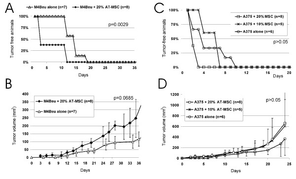
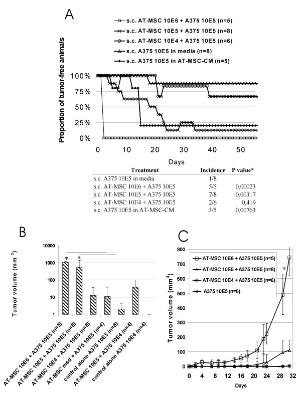
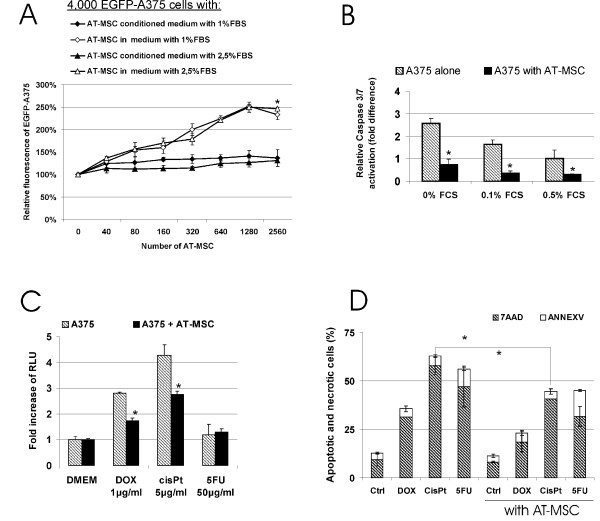
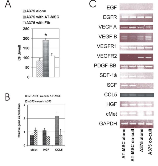
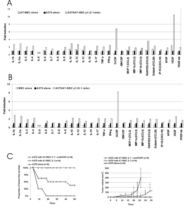
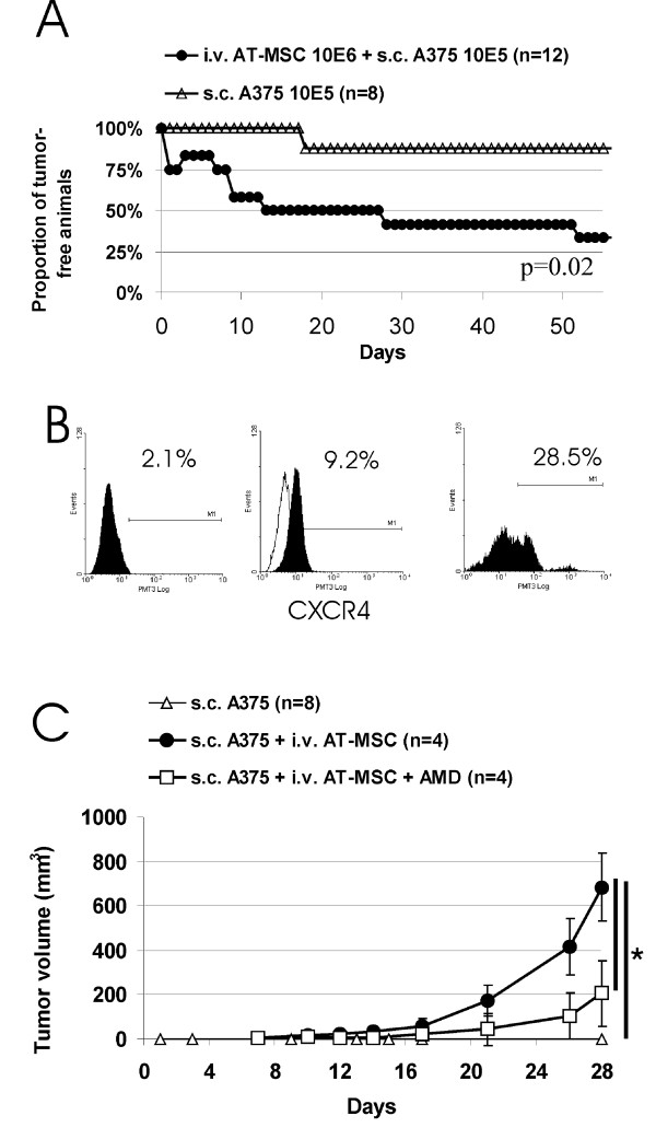
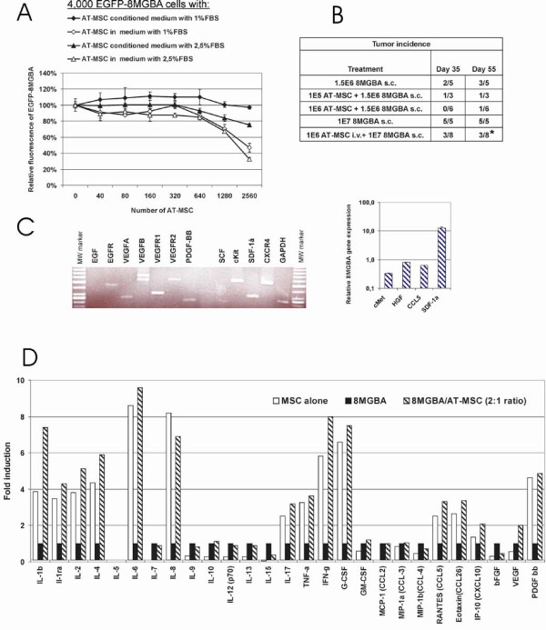
Results: Here we have demonstrated tumor-promoting effect of AT-MSC on human melanoma A375 cells. AT-MSC coinjection mediated abrogation of tumor latency and supported subcutaneous xenotransplant growth from very low melanoma cell doses. Tumor incidence was also significantly increased by AT-MSC-derived soluble factors. AT-MSC supported proliferation, suppressed apoptosis and modulated melanoma cell responses to cytotoxic drugs in vitro. Expression and multiplex cytokine assays confirmed synergistic increase in VEGF that contributed to the AT-MSC-mediated support of A375 xenotransplant growth. Production of G-CSF and other factors implicated in formation of supportive proinflammatory tumor cell microenvironment was also confirmed. SDF-1alpha/CXCR4 signalling contributed to tumor-promoting effect of systemic AT-MSC administration on A375 xenotransplants. However, no support was observed for human glioblastoma cells 8MGBA co-injected along with AT-MSC that did not sustain tumor xenotransplant growth in vivo. Tumor-inhibiting response could be attributed to the synergistic action of multiple cytokines produced by AT-MSC on glioblastoma cells.

Conclusions: Herein we provide experimental evidence for MSC-mediated protective effect on melanoma A375 cells under nutrient-limiting and hostile environmental conditions resulting from mutual crosstalk between neoplastic and non-malignant cells. This tumor-favouring effect was not observed for the glioblastoma cells 8MGBA. Collectively, our data further strengthen the need for unravelling mechanisms underlying MSC-mediated modulation of tumor behaviour for possible future MSC clinical use in the context of malignant disease.


Figures

Figure 1
Figure 1
AT-MSC coinjection with high melanoma cell dose changes the time of xenograft onset and growth. A.-B. 20% AT-MSC co-administered s.c. with 1 × 10⁶ M4Beu significantly shortened time to 100% tumor onset from day 19 (control) to day 12, but did not significantly increase tumor burden. C.-D. 10% or 20% AT-MSC admixed with 1.5 × 10⁶ A375 melanoma cells also shortened time of xenograft onset and increased tumor burden in nude mice.
Figure 2
Figure 2
AT-MSC abrogated tumor dormancy of low melanoma cell dose and supported tumor growth. 1 × 10⁵ or 1 × 10⁴ A375 cells resuspended in serum-free culture media were injected s.c. either alone or admixed to AT-MSC in ratios 10:1, 1:1 or 1:10. Low A375 cell dose (1 × 10⁵ s.c.) resuspended in cell-and-serum free AT-MSC-conditioned media was injected in a separate groups of animals. A. AT-MSC admixed to the A375 at a ratio 1:1 or 10:1 significantly increased tumor incidence in nude mice. AT-MSC conditioned media also abrogated tumor dormancy of low A375 melanoma dose. B. Tumor burden in 10:1 and 1:1 AT-MSC coinjected groups was significantly higher in comparison to A375 cell-induced xenografts by day 40. C. AT-MSC co-administration resulted in melanoma xenograft tumor growth support proportional to the AT-MSC cell dose (*P < 0.05).
Figure 3
Figure 3
AT-MSC increased A375 proliferation and decreased apoptosis in response to cellular stress in direct cocultures in vitro. A. Proliferation of EGFP-A375 cells when mixed with increasing numbers of AT-MSC or maintained in AT-MSC conditioned low-serum medium was evaluated by relative fluorescence after 3 days. AT-MSC significantly supported tumor cell proliferation in a dose dependent manner. This effect was significantly higher in comparison to proliferation support mediated by conditioned media from the same number of AT-MSC (*p < 0.05). B. A375 cells alone or mixed with 10% AT-MSC were maintained in 0%, 0.1% or 0.5% serum-containing medium for 48 hrs and relative Caspase-3/7 activation was evaluated by luminescence caspase assay. Caspase-3/7 activity of A375 cells in 0.5% serum-containing medium was set as 100%. AT-MSC significantly decreased caspase-3/7 activation in A375 melanoma cells. C. A375 cells alone or mixed with 10% AT-MSC were treated with doxorubicin (DOX), cisplatin (cisPt) and 5-fluorouracil (5FU) for 16 hrs under serum-deprivation conditions and Caspase 3/7 activation was evaluated by luminescence caspase assay. Results were expressed as mean increase in relative luminescence units (RLU) over background luminescence in DMEM cultured cells. AT-MSC could significantly decrease extent of caspase activation induced by doxorubicin and cisplatin in A375 cells. D. A375 cells alone or mixed with CFDA-SE-AT-MSC were treated with doxorubicin, cisplatin and 5-fluorouracil for 20 hrs. Proportion of apoptotic and necrotic A375 cells was determined by Annexin V and 7-AAD, respectively. AT-MSC decreased proportion of apoptotic and necrotic A375 cells thus reducing the cytotoxicity effect mediated by doxorubicin and cisplatin (*P < 0.05).
Figure 4
Figure 4
Paracrine signalling in indirect AT-MSC/A375 co-cultures. A. AT-MSC increased colony-formation by A375 cells. A375 cells were plated in the lower well part and combined with insert containing AT-MSC, fibroblast or no cells in upper part for 9 days in indirect coculture in vitro. Average number of A375 colonies/bottom well is shown in one representative experiment performed in triplicates (*P < 0.05). B. Quantitative RT-PCR was performed on templates isolated from indirectly cocultured A375 and AT-MSC cells. Gene expression level was compared to parental cells and expressed as relative gene expression. HGF and cMet expression is sustained upon coculture, CCL5 increases in AT-MSC cells cocultured with A375 cells for 3 days. C. Semi-quantitative expression analysis of parental A375, AT-MSC and cocultured A375 and AT-MSC cells confirms sustained expression of EGFR, VEGF A, VEGF B, VEGFR1, PDGF-bb, SDF-1α, SCF, CCL5, HGF, cMet in AT-MSC; and EGFR, VEGF-A,-B, VEGFR-1,-2 PDGF-bb, SDF-1α, CCL5, HGF, cMet in A375.
Figure 5
Figure 5
Direct AT-MSC/A375 interactions synergistically increase VEGF production and its neutralization can partially inhibit AT-MSC mediated tumor onset and growth support. A375, AT-MSC, or A375/AT-MSC were directly co-cultured in complete media for 3 days. A375/AT-MSC ratio was A) 2:1 or B) 5:1. The level of cytokines in cell-free supernatants was determined by Bio-Plex cytokine arrays and normalized to the levels observed in the media of A375 cell cultured alone. Mostly, the effects were additive or slightly synergistic. The levels of G-CSF and VEGF were significantly increased in both cases and exhibited potent synergistic effect. Data were expressed as average fold induction. ND, not detected. C) 2 × 10⁵ A375 cells resuspended in serum-free culture media were injected s.c. either alone or admixed to AT-MSC in ratio 2:1. One group of animals was treated with 1 mg/kg Avastin twice a week as indicated (antiVEGF group). Left panel: AT-MSC admixed to the A375 at a ratio 2:1 significantly increase tumor growth in nude mice and abrogate tumor dormancy (p = 0.0013). VEGF neutralization significantly inhibited protumorigenic AT-MSC mediated effect (p = 0.0498). Right panel: AntiVEGF treatment significantly decreased tumor burden and counteracted protumorigenic effects of AT-MSC (*p < 0.05).
Figure 6
Figure 6
Systemic AT-MSC administration abrogates A375 tumor dormancy and AT-MSC tumor growth support can be partially blocked by inhibiting SDF-1α/CXCR4 signalling. A. Systemic administration of AT-MSC (106 i.v.) concomitant with the implantation of 1 × 10⁵ A375 s.c. resulted in abrogation of tumor dormancy in 8 out of 12 cases in contrast to 1 out of 8 implantations of A375 s.c. alone. B. Cultured A375 cells or single-cell suspensions prepared by positive immunomagnetic separation of human CD44+ cells from tumor xenotransplants were stained with anti-CXCR4 antibody. Flow cytometric analysis has shown the absence of the CXCR4 marker on low density cultured A375 cells (left), CXCR4 increase upon cell confluence (middle) and high level of expression on A375 from tumor xenotransplant in vivo (right). CXCR4 (filled area), isotype control (open area). C. 2 × 10⁵ A375 were implanted s.c either alone or coimplanted with AT-MSC (106 i.v.). Although all xenografts in AT-MSC injected group started to grow, animals treated with AMD3100 inhibitor of SDF-1α/CXCR4 (1.25 mg/kg every other day s.c.) exhibited significantly lower tumor volume in comparison to untreated group (*p < 0.05).
Figure 7
Figure 7
8MGBA glioblastoma proliferation and tumor incidence was not increased by AT-MSC. A. Proliferation of EGFP-8MGBA cells when admixed with increasing numbers of AT-MSC or maintained in AT-MSC conditioned low-serum medium was evaluated by relative fluorescence after 3 days. AT-MSC did not support tumor cell proliferation in comparison to control without AT-MSC. EGFP-8MGBA proliferation was significantly inhibited in co-culture containing 39% of AT-MSC (*p < 0.05). B. 8MGBA cells 1.5 × 10⁶ or 1 × 10⁷ were injected s.c. either alone, admixed to AT-MSC at a ratio 1:1 or 1:10 or 1 × 10⁶ AT-MSC i.v. AT-MSC decreased the tumor incidence by day 55 from 60% in 8MGBA alone group to 33% in 8MGBA/AT-MSC 10:1 group and 17% in 8MGBA/AT-MSC 1:1 group. Systemic AT-MSC administration significantly decreased tumor incidence to 37.5% (*p = 0.0304). C. 8MGBA expression profile demonstrated expression of EGFR, VEGF-A,-B, VEGFR-1,-2 PDGF-bb, cKit, SDF-1α (high), CXCR4, CCL5, HGF, cMet (low). Quantitative differences were detected in higher level of SDF-1α expression and lower level of cMet receptor expression in comparison to A375 melanoma. D. 8MGBA, AT-MSC, or 8MGBA/AT-MSC (ratio 2:1) were directly co-cultured in complete media for 3 days. Level of cytokines in cell-free supernatants was determined by Bio-Plex cytokine arrays and normalized to the levels observed in the media of 8MGBA cells cultured alone. Mostly, the effects were additive, increased level of IL-1β and IFN-γ was observed in directly cocultured cells. Data were expressed as average fold induction. ND, not detected.

References

    1. Giordano A, Galderisi U, Marino IR. From the laboratory bench to the patient's bedside: an update on clinical trials with mesenchymal stem cells. J Cell Physiol. 2007;211:27–35. doi: 10.1002/jcp.20959. - DOI - PubMed
    1. Le Blanc K, Ringdén O. Immunomodulation by mesenchymal stem cells and clinical experience. J Intern Med. 2007;262:509–25. doi: 10.1111/j.1365-2796.2007.01844.x. - DOI - PubMed
    1. Picinich SC, Mishra PJ, Mishra PJ, Glod J, Banerjee D. The therapeutic potential of mesenchymal stem cells. Cell- & tissue-based therapy. Expert Opin Biol Ther. 2007;7:965–73. doi: 10.1517/14712598.7.7.965. - DOI - PubMed
    1. Psaltis PJ, Zannettino AC, Worthley SG, Gronthos S. Concise review: mesenchymal stromal cells: potential for cardiovascular repair. Stem Cells. 2008;26:2201–10. doi: 10.1634/stemcells.2008-0428. - DOI - PubMed
    1. Uccelli A, Moretta L, Pistoia V. Mesenchymal stem cells in health and disease. Nat Rev Immunol. 2008;8:726–36. doi: 10.1038/nri2395. - DOI - PubMed

Publication types

LinkOut - more resources