

Canine classical seminoma: a specific malignant type with human classifications is highly correlated with tumor angiogenesis
- PMID: 20509912
- PMCID: PMC2887404
- DOI: 10.1186/1471-2407-10-243
Canine classical seminoma: a specific malignant type with human classifications is highly correlated with tumor angiogenesis
Abstract
Background: Human seminoma is classified as classical seminoma (SE) and spermatocytic seminoma (SS). Human SE is known to be more malignant and metastasizing more frequently than SS. Tumor angiogenesis is highly related with tumor progression and metastasis, with microvessel density (MVD) being an important parameter of metastatic potential. Canine seminoma is not yet well-established as SE or SS type including correlation with angiogenesis. We classified canine SE and SS, and then compared them to tumor associated vessels.
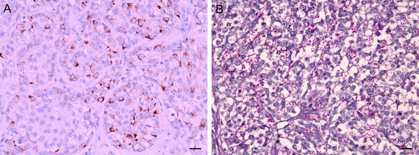
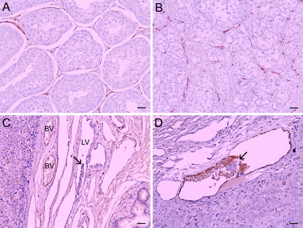
Methods: Twenty-three cases of canine seminomas (2 intratubular, 9 diffuse, and 12 intratubular/diffuse seminomas showing both intratubular and diffuse patterns) were classified as SE or SS by immunohistochemistry (IHC) using monoclonal antibody against PLAP and by PAS stain. The histopathological data were then compared to see if there was a correlation with SE or SS. Angiogenesis of seminomas were evaluated by immunohistochemical assay using polyclonal antibody against Von Willebrand factor (vWF) and by calculating the means of MVD, vessels area and perimeters using computerized image analysis. Statistical Package for Social Sciences (SPSS) program was used for various statistical analyses.
Results: The numbers of PLAP+/PAS+ canine SEs were 8/23 (34.8%) and PLAP-/PAS- SSs were 15/23 (61.2%). All SE cases (8/8, 100%) were intratubular/diffuse types. SS types included 2 intratubular (2/15, 13.3%), 9 diffuse (9/15, 60%), and 4 intratubular/diffuse (4/15, 26.7%) types. MVD and vascular parameters in SEs were significantly higher than in SSs, showing the highest value in the intratubular/diffuse type. Seminomas observed with neoplastic cells invasion of vessels presented higher perimeter and area values than seminomas without conformed neoplastic cells invasion.
Conclusion: In this study, we demonstrated a positive relationship between canine SE and tumor angiogenesis. Furthermore, we also showed that a tumor cells invasion of vessels were a correlated vascular parameter. Although metastasis of canine seminomas has rarely been reported, our results support that canine SE could have high metastatic potential similar to the human counterpart. Further studies are required to clarify the relationship between canine SE and clinical data with metastatic factors.
Figures
Similar articles
-
Evaluation of angiogenesis in canine seminomas by quantitative immunohistochemistry.J Comp Pathol. 2003 May;128(4):252-9. doi: 10.1053/jcpa.2002.0630. J Comp Pathol. 2003. PMID: 12834608
-
Classical and spermatocytic seminoma in the dog: histochemical and immunohistochemical findings.J Comp Pathol. 2007 Jul;137(1):41-46. doi: 10.1016/j.jcpa.2007.03.009. J Comp Pathol. 2007. PMID: 17629966
-
Expression of protein gene product 9.5 and Sal-like protein 4 in canine seminomas.J Comp Pathol. 2014 Jul;151(1):10-8. doi: 10.1016/j.jcpa.2014.02.003. Epub 2014 Feb 12. J Comp Pathol. 2014. PMID: 24680979
-
Correlation of nuclear morphometric features with animal and human World Health Organization International Histological Classifications of canine spontaneous seminomas.Vet Pathol. 2004 Nov;41(6):608-11. doi: 10.1354/vp.41-6-608. Vet Pathol. 2004. PMID: 15557070
-
[Spermatocytic seminoma. A tumor with many faces].Pathologe. 2014 May;35(3):232-7. doi: 10.1007/s00292-014-1899-x. Pathologe. 2014. PMID: 24682373 Review. German.
Cited by
-
Morphological and immunohistochemical characterisation of seminomas in Norwegian dogs.Acta Vet Scand. 2012 Sep 17;54(1):52. doi: 10.1186/1751-0147-54-52. Acta Vet Scand. 2012. PMID: 22986090 Free PMC article.
-
Correlation of tumor-infiltrating lymphocytes to histopathological features and molecular phenotypes in canine mammary carcinoma: A morphologic and immunohistochemical morphometric study.Can J Vet Res. 2013 Apr;77(2):142-9. Can J Vet Res. 2013. PMID: 24082407 Free PMC article.
-
Canine testicular tumors: two types of seminomas can be differentiated by immunohistochemistry.BMC Vet Res. 2014 Aug 6;10:169. doi: 10.1186/s12917-014-0169-8. BMC Vet Res. 2014. PMID: 25096628 Free PMC article.
-
The roles and mechanisms of hypoxia in liver fibrosis.J Transl Med. 2021 May 1;19(1):186. doi: 10.1186/s12967-021-02854-x. J Transl Med. 2021. PMID: 33933107 Free PMC article. Review.
-
Immunophenotyping of Rabbit Testicular Germ and Sertoli Cells Across Maturational Stages.J Histochem Cytochem. 2016 Nov;64(11):715-726. doi: 10.1369/0022155416669918. Epub 2016 Sep 30. J Histochem Cytochem. 2016. PMID: 27680667 Free PMC article.
References
-
- Ulbright TM, Roth LM. Recent developments in the pathology of germ cell tumors. Semin Diagn Pathol. 1987;4(4):304–319. - PubMed
Publication types
MeSH terms
Substances
LinkOut - more resources
Full Text Sources
Medical
Miscellaneous
