. 2010;12(3):R106.
doi: 10.1186/ar3039. Epub 2010 May 28.

Mechanical signals control SOX-9, VEGF, and c-Myc expression and cell proliferation during inflammation via integrin-linked kinase, B-Raf, and ERK1/2-dependent signaling in articular chondrocytes

Affiliations

Mechanical signals control SOX-9, VEGF, and c-Myc expression and cell proliferation during inflammation via integrin-linked kinase, B-Raf, and ERK1/2-dependent signaling in articular chondrocytes

Priyangi M Perera et al. Arthritis Res Ther. 2010.

Abstract

Introduction: The importance of mechanical signals in normal and inflamed cartilage is well established. Chondrocytes respond to changes in the levels of proinflammatory cytokines and mechanical signals during inflammation. Cytokines like interleukin (IL)-1beta suppress homeostatic mechanisms and inhibit cartilage repair and cell proliferation. However, matrix synthesis and chondrocyte (AC) proliferation are upregulated by the physiological levels of mechanical forces. In this study, we investigated intracellular mechanisms underlying reparative actions of mechanical signals during inflammation.

Methods: ACs isolated from articular cartilage were exposed to low/physiologic levels of dynamic strain in the presence of IL-1beta. The cell extracts were probed for differential activation/inhibition of the extracellular signal-regulated kinase 1/2 (ERK1/2) signaling cascade. The regulation of gene transcription was examined by real-time polymerase chain reaction.

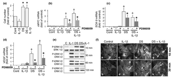
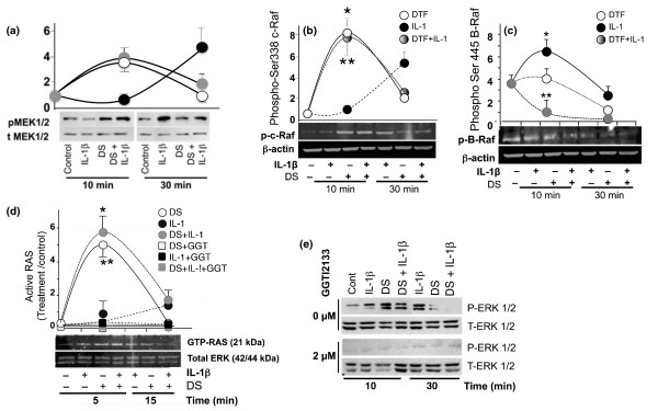
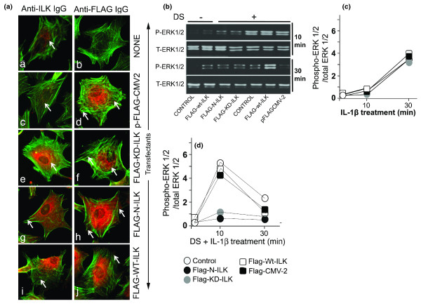
Results: Mechanoactivation, but not IL-1beta treatment, of ACs initiated integrin-linked kinase activation. Mechanical signals induced activation and subsequent C-Raf-mediated activation of MAP kinases (MEK1/2). However, IL-1beta activated B-Raf kinase activity. Dynamic strain did not induce B-Raf activation but instead inhibited IL-1beta-induced B-Raf activation. Both mechanical signals and IL-1beta induced ERK1/2 phosphorylation but discrete gene expression. ERK1/2 activation by mechanical forces induced SRY-related protein-9 (SOX-9), vascular endothelial cell growth factor (VEGF), and c-Myc mRNA expression and AC proliferation. However, IL-1beta did not induce SOX-9, VEGF, and c-Myc gene expression and inhibited AC cell proliferation. More importantly, SOX-9, VEGF, and Myc gene transcription and AC proliferation induced by mechanical signals were sustained in the presence of IL-1beta.

Conclusions: The findings suggest that mechanical signals may sustain their effects in proinflammatory environments by regulating key molecules in the MAP kinase signaling cascade. Furthermore, the findings point to the potential of mechanosignaling in cartilage repair during inflammation.


Figures

Figure 1
Figure 1
Mechanical signals upregulate articular chondrocyte (AC) proliferation via SOX-9, VEGF, and c-Myc mRNA expression and ERK1/2 activation. ACs were exposed to no treatment or to treatment with interleukin-1-beta (IL-1β), dynamic strain (DS) alone, or DS and IL-1β. Subsequently, ACs were subjected to DS for 90 minutes per day for 3 days. (a) On day 4, the rate of cell proliferation was assessed by MTT assay. ACs were treated either with medium alone or with PD98059 (2 μM) for 30 minutes. Cells were exposed to the treatment regimens above for 3 hours, and the mRNA expression for c-Myc (b), SOX-9 (c), and VEGF (d) was analyzed by real-time polymerase chain reaction. (e) Western blot analysis showing ERK1/2 phosphorylation using phospho-Thr202/Tyr204 ERK1/2 (P-ERK1/2) and total ERK1/2 (T-ERK1/2) antibodies. (f) Immunofluorescence analysis showing minimal phospho-ERK1/2 in control cells [a], cells stained with secondary antibody alone [b], optimal phosphorylation of ERK1/2 and its nuclear translocation in response to IL-1β at 10 and 30 minutes [c,d], and nuclear translocation and cytoplasmic redistribution of p-ERK1/2 in response to DS in the absence [e,f] and presence [g,h] of IL-1β. Cells were counterstained with fluorescein isothiocyanate-phalloidin to show β-actin. Experiments in (a,c-e) were performed in triplicate and were repeated three times in (b) and two times in (f). The error bars represent standard error of the mean (standard error of the mean in a-d). Gels in (e) represent one of three experiments with similar results. Φ P < 0.05 as compared with untreated controls; *P < 0.05 as compared with cells treated with DS or with DS and IL-1. C, control; Cont, control; ERK1/2, extracellular receptor kinase 1/2; MTT, 3-(4,5 dimethylthiazolyl-2)-2,5-diphenyltetrazolium bromide; SOX-9, SRY-related protein-9; VEGF, vascular endothelial cell growth factor.
Figure 2
Figure 2
Mitogen-activated protein kinase signaling in response to dynamic strain (DS). Articular chondrocytes (ACs) were exposed to no treatment or to treatment with interleukin-1-beta (IL-1β), DS alone, or DS and IL-1β for 10 or 30 minutes and were examined by Western blot analysis for (a) Ser217/221-MEK1/2 phosphorylation by DS and IL-1β. Equal protein loading was confirmed by probing blots with anti-total MEK1/2 antibody. (b) Ser338-c-RAF phosphorylation, (c) Ser445-B-Raf phosphorylation, and (d) Ras activation following immunoprecipitation with GST-Raf-1-RBD and glutathione agarose beads are shown. (e) Ras activation following pretreatment of cells with a selective Ras antagonist (2 μM GGT12133) for 30 minutes is shown along with the assessment of Ras-dependent phospho-Thr202/Tyr204-ERK1/2 (P-ERK1/2) 10 and 30 minutes post-activation. Anti-total ERK1/2 IgG (T-ERK1/2) was used to normalize total input in all lanes. All experiments were performed in triplicate. Gels represent one of three experiments with similar results in (a-e). The graphs above gels in each figure show mean and standard error of the mean of phosphoprotein/total protein in three separate experiments in (A-D). *P < 0.05 as compared with untreated control cells; **P < 0.05 as compared with IL-1β-treated cells. DTF, dynamic tensile force; ERK1/2, extracellular receptor kinase 1/2; GGT, Ras inhibitor GGT12133; GST, glutathione-S-transferase; MEK1/2, mitogen-activated protein kinase/extracellular receptor kinase 1/2; RBD, Ras-binding domain.
Figure 3
Figure 3
Mechanical signals activate integrin-linked kinase (ILK). (a) Articular chondrocytes (ACs) either were not transfected [a,b] or were transfected with p-FLAG-CMV2 empty [c,d], FLAG-KD-ILK [e,f], FLAG-N-ILK [g,h], or FLAG-WT ILK [i,j]. ACs were immunostained with anti-ILK (left frames) or anti-FLAG (right frames) antibodies and CY3-conjugated secondary antibodies. All cells were counterstained with fluorescein isothiocyanate-phalloidin to visualize β-actin. Western blot analysis shows ERK1/2 activation in untransfected ACs or those transfected with FLAG-N-ILK, FLAG-KD-ILK, FLAG-WT-ILK, or pFLAG-CMV2 exposed to (b) no strain or dynamic strain (DS) alone, (c) interleukin-1-beta (IL-1β) alone, or (d) DS and IL-1β. Frames (c,d) show semiquantitative estimation of bands in Western blots. All figures represent one of three similar experiments. ERK1/2, extracellular receptor kinase 1/2; FLAG, polypeptide protein sequence DYKDDDDK; KD, kinase-deficient; P-ERK1/2, phospho-Thr202/Tyr204-extracellular receptor kinase 1/2; T-ERK1/2, total extracellular receptor kinase 1/2; WT, wild-type.

Similar articles

Cited by

References

    1. Zuscik MJ, Hilton MJ, Zhang X, Chen D, O'Keefe RJ. Regulation of chondrogenesis and chondrocyte differentiation by stress. J Clin Invest. 2008;118:429–438. doi: 10.1172/JCI34174. - DOI - PMC - PubMed
    1. Chiquet M, Gelman L, Lutz R, Maier S. From mechanotransduction to extracellular matrix gene expression in fibroblasts. Biochim Biophys Acta. 2009;1793:911–920. doi: 10.1016/j.bbamcr.2009.01.012. - DOI - PubMed
    1. Dossumbekova A, Anghelina M, Madhavan S, He L, Quan N, Knobloch T, Agarwal S. Biomechanical signals inhibit IKK activity to attenuate NF-kappaB transcription activity in inflamed chondrocytes. Arthritis Rheum. 2007;56:3284–3296. doi: 10.1002/art.22933. - DOI - PMC - PubMed
    1. Agarwal S, Deschner J, Long P, Verma A, Hofman C, Evans CH, Piesco N. Role of NF-kappaB transcription factors in antiinflammatory and proinflammatory actions of mechanical signals. Arthritis Rheum. 2004;50:3541–3548. doi: 10.1002/art.20601. - DOI - PMC - PubMed
    1. Wang B, Yang Y, Friedman PA. Na/H exchange regulatory factor 1, a novel AKT-associating protein, regulates extracellular signal-regulated kinase signaling through a B-raf-mediated pathway. Mol Biol Cell. 2008;19:1637–1645. doi: 10.1091/mbc.E07-11-1114. - DOI - PMC - PubMed

Publication types

MeSH terms

Substances