Comparative Study
. 2010 Aug 15;344(2):1071-87.
doi: 10.1016/j.ydbio.2010.05.495. Epub 2010 May 24.

Comparative gene expression analysis of genital tubercle development reveals a putative appendicular Wnt7 network for the epidermal differentiation

Affiliations
Comparative Study

Comparative gene expression analysis of genital tubercle development reveals a putative appendicular Wnt7 network for the epidermal differentiation

Han Sheng Chiu et al. Dev Biol. .

Abstract

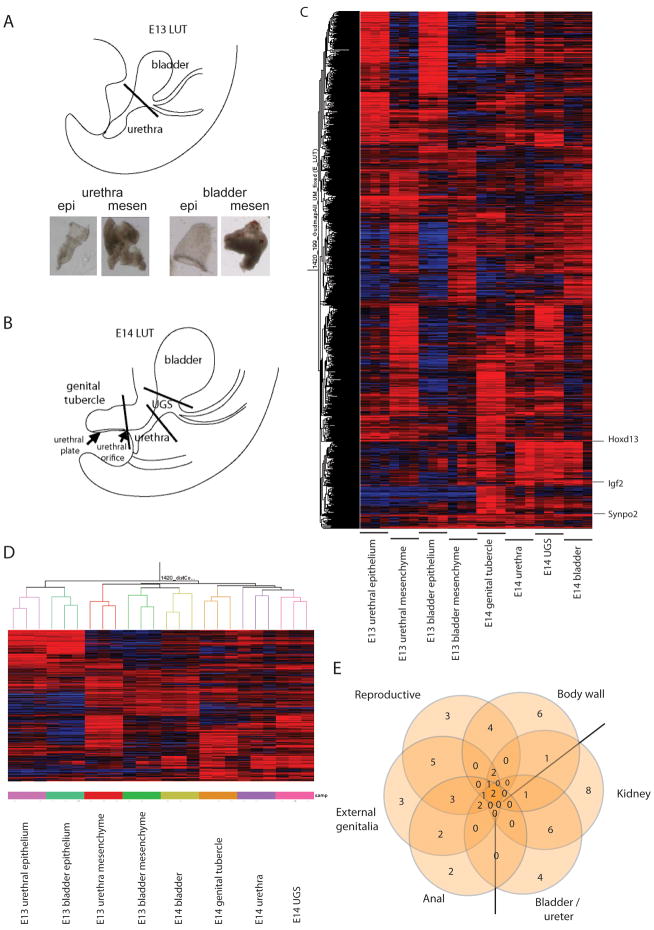
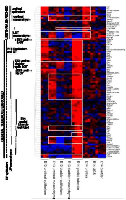
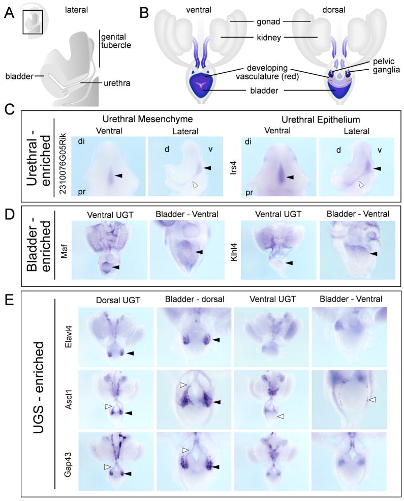
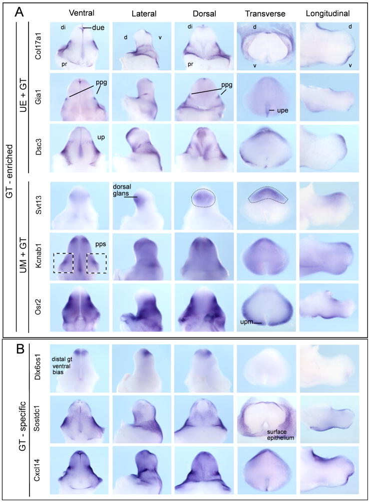
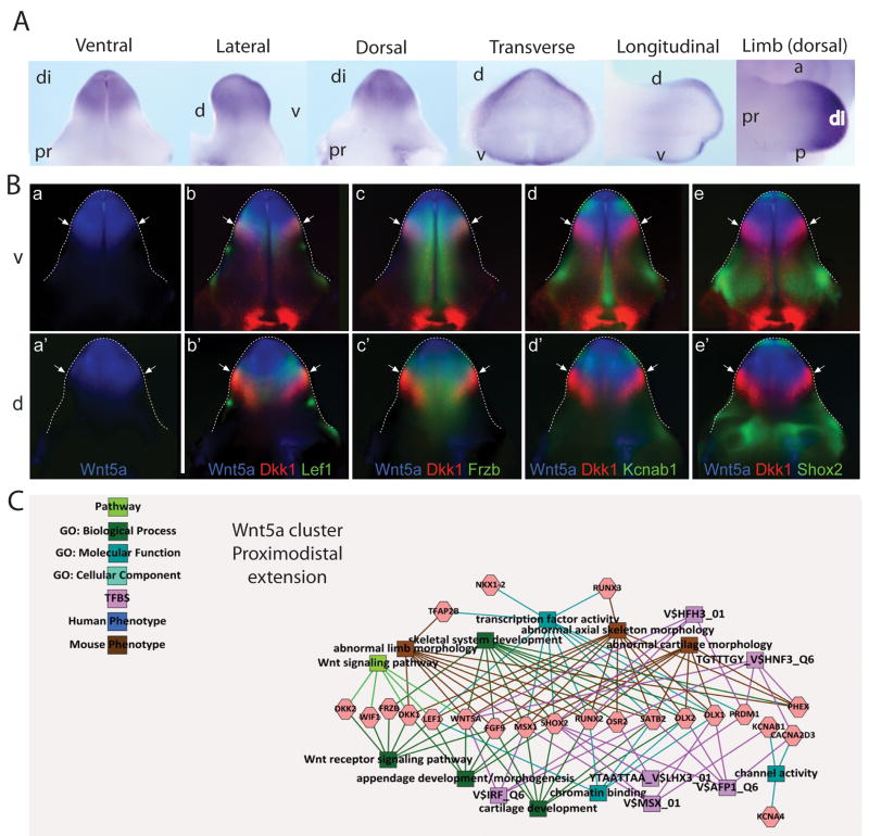
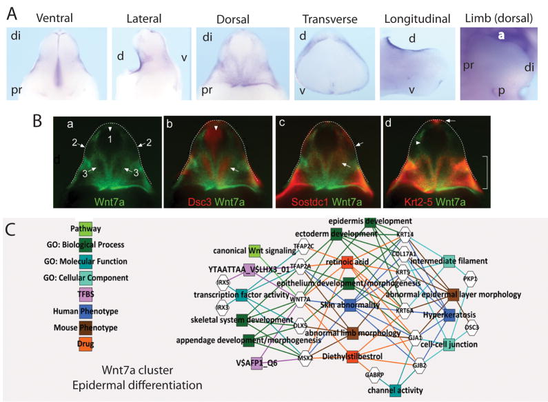
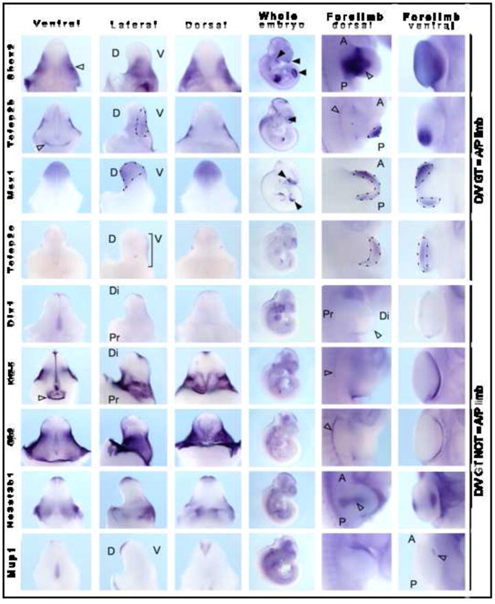
Here we describe the first detailed catalog of gene expression in the developing lower urinary tract (LUT), including epithelial and mesenchymal portions of the developing bladder, urogenital sinus, urethra, and genital tubercle (GT) at E13 and E14. Top compartment-specific genes implicated by the microarray data were validated using whole-mount in situ hybridization (ISH) over the entire LUT. To demonstrate the potential of this resource to implicate developmentally critical features, we focused on gene expression patterns and pathways in the sexually indeterminate, androgen-independent GT. GT expression patterns reinforced the proposed similarities between development of GT, limb, and craniofacial prominences. Comparison of spatial expression patterns predicted a network of Wnt7a-associated GT-enriched epithelial genes, including Gjb2, Dsc3, Krt5, and Sostdc1. Known from other contexts, these genes are associated with normal epidermal differentiation, with disruptions in Dsc3 and Gjb2 showing palmo-plantar keratoderma in the limb. We propose that this gene network contributes to normal foreskin, scrotum, and labial development. As several of these genes are known to be regulated by, or contain cis elements responsive to retinoic acid, estrogen, or androgen, this implicates this pathway in the later androgen-dependent development of the GT.


Figures

Figure 1
Figure 1. Defining the gene expression profile of the developing lower urinary tract
A) Diagram of the E13 LUT indicating the point of dissection to separate E13 urethra from E13 bladder and the resultant separation of epithelium from mesenchyme achieved using trypsin digestion and EDTA dissociation, respectively. B) Diagram of the E14 LUT indicating the point of dissection to delineate four spatial regions; genital tubercle (GT), urethra, UGS and bladder. C) Heat map of the 1420 most highly expressed genes across all eight compartments of the developing LUT. D) Cluster diagram highlighting the hierarchical relationships between all samples analysed (eight compartments in triplicate). Note that all replicates correctly clustered with their compartment of origin. E) Venn diagram indicating the degree of gene overlap between the processes of reproductive, external genital, anal, body wall, kidney and bladder/ureter development identified in the GT dataset.
Figure 2
Figure 2. Heat map of gene expression across E14 urethral-enriched and genital tubercle-enriched genes
Urethral-enriched genes can be subdivided into epithelial versus mesenchymal based on the expression in E13 urethra. Genital tubercle-enriched genes were subdivided into urethral plate (UP) epithelium (expressed in genital tubercle and E13 urethral plate epithelium but not E14 urethra), UP mesenchyme (expressed in genital tubercle and E13 urethral mesenchyme but not E14 urethra) and genital tubercle-specific genes (expressed only in GT).
Figure 3
Figure 3. Validation of E14 urethral, bladder and UGS-enriched genes
A) Schematic diagram of a bisected LUT/GT, lateral view, illustrating the bladder, UGS and urethra. up, urethral plate; due, distal urethral epithelium B) Schematic diagram of whole urogenital tracts, ventral and dorsal views. C) Urethral-enriched genes with epithelial (lrs4) and mesenchymal (2310076G05Rik) expression displayed as ventral (left) and lateral (right) views. Expression can be seen along the UP (solid arrowheads) and in the urethra itself (open arrowheads). D) E13 bladder-enriched genes Maf and Klhl4 showing expression in vasculature and differential expression in the dome of the bladder (solid arrowheads, rostral end). E) UGS-enriched genes Elav4, Ascl1 and Gap43 revealing specific expression in migrating neural crest cells (open arrowheads) and developing ganglia (solid arrowheads) at the base of the dorsal surface of the bladder. di, distal; pr, proximal; d, dorsal; v, ventral.
Figure 4
Figure 4. Validation of E14 genital tubercle-enriched gene expression
A) Genital tubercle-enriched genes predicted to be expressed in UP epithelium and GT (Col17a, Gja1, Dsc3) or UP mesenchyme and GT (Syt13, Kcnab1, Osr2). Expression can also be seen in preputial gland (Gja1), distal urethral epithelium (Col17a, Gja1, Dsc3), surface epithelium of glans and preputial swelling (Col17a, Gja1, Dsc3) and preputial swelling mesenchyme (Kcnab1, Osr2). B) Genital tubercle-restricted genes (Dlx6os1, Sostdc1, Cxcl14) showed expression in preputial swelling epithelium (Sostdc1, Cxcl14), distinct domains across the dorsal GT (Dlx6os1, Sostdc1, Cxcl14) and distal genital tubercle (glans) (Dlx6os1). Material is presented from left to right as wholemounts of ventral GT, lateral GT (dorsal to the left side), dorsal GT, and transverse and longitudinal sections through wholemount specimens. di, distal; pr, proximal; d, dorsal; v, ventral; due, distal urethral epithelium; up, urethral plate; upe, up epithelium; upm, up mesenchyme; ppg, preputial gland; pps, preputial swelling.
Figure 5
Figure 5. Identification of Wnt5a associated gene networks in GT development
A. Wholemount ISH of Wnt5a in the developing E13.5 GT showing ventral, lateral, dorsal, transverse and longitudinal views, as well as Wnt5a expression in the limb (dorsal view). di, distal; pr, proximal; d, dorsal; v, ventral; a, anterior; p, posterior. B. Pseudo-colored expression pattern overlays of known and candidate genes of a Wnt5a network. Experimental WISH images of stage-matched genital tubercles at 13.5dpc were pseudo-colored and overlaid with either Wnt5a (a–e, a′–e′). Ventral (v; a–e) and dorsal (d; a′–e′) views are shown and the edge of the GT is outlined with a dotted line. Wnt5a network genes include Wnt5a alone (a, a′), Wnt5a/Dkk1/Lef1 (b, b′), Wnt5a/Dkk1/Frzb (c, c′), Wnt5a/Dkk1/Kcnab1 (d, d′) and Wnt5a/Dkk1/Shox2 (e, e′). Arrows indicate the border of the Wnt5a distal GT expression domain. All genes showed some degree of overlap with the Wnt5a domain. Dkk1 showed overlapping expression with Lef1 (b, b′) and Frzb (c, c′), in contrast to Kcnab1 (d, d′) and Shox2 (e, e′), which did not overlap with the distal Dkk1 domain. C. GT mesenchyme-enriched gene cluster associated via hierarchical clustering decorated with GO terms and known transcription factor binding sites and known human and mouse phenotype associations. This cluster shows strong links to limb morphogenesis, particularly of mesodermal elements including cartilage and bone.
Figure 6
Figure 6. Identification of Wnt7a associated gene networks in GT development
A. Wholemount ISH of Wnt7a in the developing E13.5 GT showing ventral, lateral, dorsal, transverse and longitudinal views, as well as Wnt5a expression in the limb (dorsal view). B. Pseudo-colored expression pattern overlays of known and candidate genes of a Wnt7a network. Experimental WISH images of stage-matched genital tubercles at 13.5dpc were pseudo-colored and overlaid with Wnt7a (a–d). Dorsal (d; a–d) views are shown and the edge of the GT is outlined with a dotted line. Wnt7a network genes include Wnt7a alone (a), Dsc3/Wnt7a (b), Sostdc1/Wnt7a (c) and Krt5/Wnt7a (d). (a) Arrows indicate the main regions (1–3) on the dorsal side of the GT where Wnt7a was not expressed. (b) Dsc3 showed a complementary pattern to Wnt7a with strong, tightly restricted expression in regions 1 and 3 (arrows), but was expressed in the epithelium. (c,d) Sostdc1 and Krt5 showed overlapping expression with Wnt7a. Sostdc1 was expressed more broadly than Wnt7a and extended into regions 2 and 3 (arrows), however like Wnt7a, expression was absent from the central region 1. (d) Krt5 expression closely resembled Wnt7a with strong expression in the proximal Wnt7a domains. In the distal Wnt7a domains, only weak Krt5 expression was seen (arrowhead), except for a small region of strong expression at the distal GT tip (arrow). C. GT epithelium-enriched gene cluster associated via hierarchical clustering decorated with GO terms and known transcription factor binding sites and known human and mouse phenotype associations. This cluster shows strong links to retinoic acid and diethylstilbestrol regulation as well as epithelium and epidermal differentiation, notably in the limbs.
Figure 7
Figure 7. Examination of the congruence between gene expression polarity in genital tubercle versus limb
Wholemount ISH was performed on E13.5 genital tubercle, E10.5 whole embryo and E10.5 forelimb. Specimens are presented from left to right as ventral GT, lateral GT, dorsal GT, E10.5 whole embryo, E11.5 dorsal limb and apical/ventral forelimb limb views. Expression of Shox2, Tcfap2b and Msx1 showed congruence of dorsal/ventral GT and anterior/posterior limb (lateral GT = DV limb). Shox2 was expressed in the mesenchyme of the paired lateral preputial swellings (open arrowhead) and was also expressed in central mesenchyme of the developing limb. Tcfap2b showed preputial swelling expression. D, dorsal; V, ventral; A, anterior; P, posterior; Di, distal; Pr, proximal.

Similar articles

Cited by

References

    1. Aboushwareb T, Zhou G, Deng FM, Turner C, Andersson KE, Tar M, Zhao W, Melman A, D’Agostino R, Jr, Sun TT, Christ GJ. Alterations in bladder function associated with urothelial defects in uroplakin II and IIIa knockout mice. Neurourol Urodyn. 2009 Epub ahead of print. - PMC - PubMed
    1. Adamska M, MacDonald BT, Meisler MH. Doubleridge, a mouse mutant with defective compaction of the apical ectodermal ridge and normal dorsal-ventral patterning of the limb. Dev Biol. 2003;255:350–362. - PubMed
    1. Ahn K, Mishina Y, Hanks MC, Behringer RR, Crenshaw EB., III BMPR-1A signaling is required for the formation of the apical ectodermal ridge and dorsal-ventral patterning of the limb. Development. 2001;128:4449–4451. - PubMed
    1. Al-Qattan MM, Al-Balwi M, Eyaid W, Al-Abdulkarim I, Al-Turki S. Congenital duplication of the palm syndrome: gene analysis and the molecular basis of its clinical features. J Hand Surgery. 2009;34E:247–251. - PubMed
    1. Beleza-Meireles A, Lundberg F, Lagerstedt K, Zhou X, Omrani D, Frisen L, Norddenskjold A. FGFR2, FGF8, FGF10 and BMP7 as candidate genes for hypospadias. Eur J Hum Genetics. 2007;15:405–410. - PubMed

Publication types