

The immunological functions of saposins
- PMID: 20510729
- PMCID: PMC4030616
- DOI: 10.1016/S0065-2776(10)05002-9
The immunological functions of saposins
Abstract
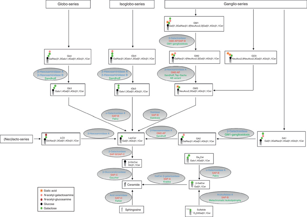
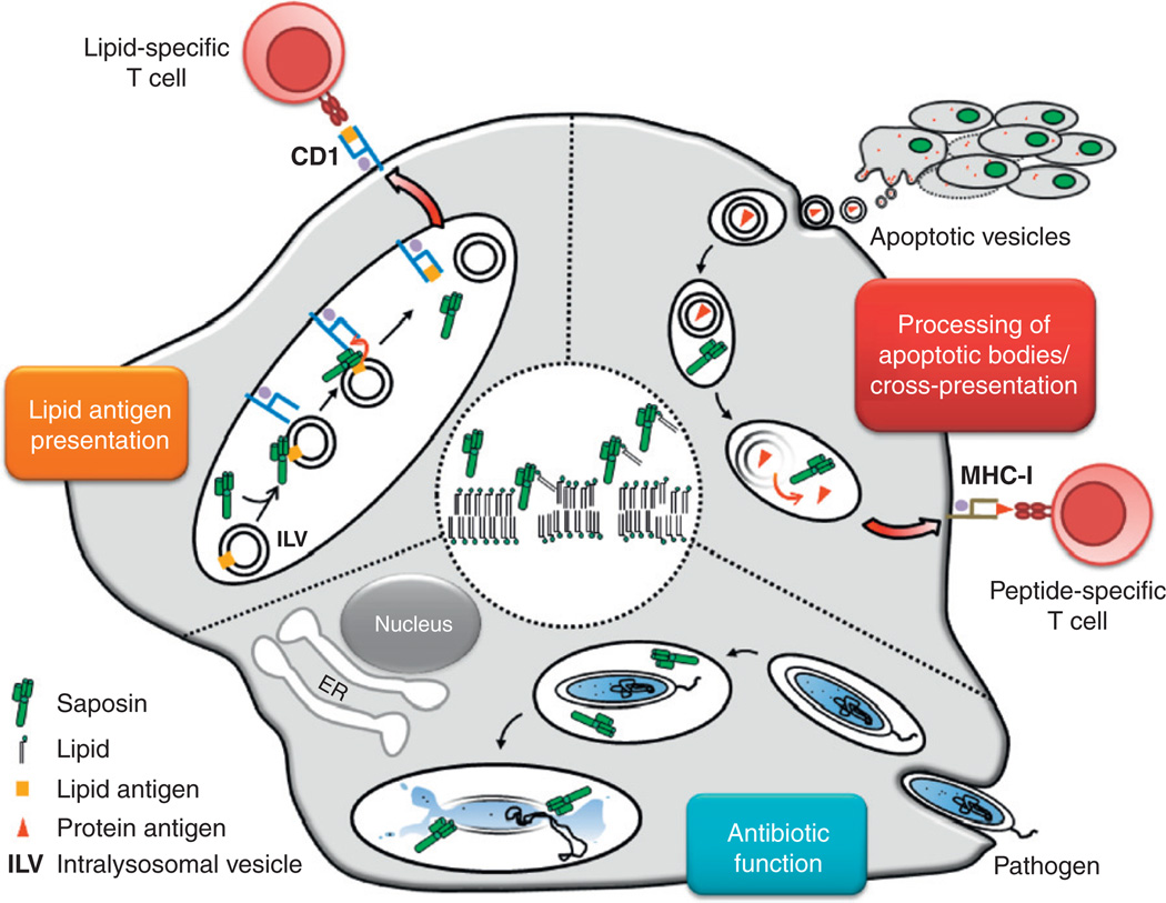
Saposins or sphingolipid activator proteins (SAPs) are small, nonenzymatic glycoproteins that are ubiquitously present in lysosomes. SAPs comprise the five molecules saposins A-D and the GM2 activator protein. Saposins are essential for sphingolipid degradation and membrane digestion. On the one hand, they bind the respective hydrolases required to catabolize sphingolipid molecules; on the other hand, saposins can interact with intralysosomal membrane structures to render lipids accessible to their degrading enzymes. Thus, saposins bridge the physicochemical gap between lipid substrate and hydrophilic hydrolases. Accordingly, defects in saposin function can lead to lysosomal lipid accumulation. In addition to their specific functions in sphingolipid metabolism, saposins have membrane-perturbing properties. At the low pH of lysosomes, saposins get protonated and exhibit a high binding affinity for anionic phospholipids. Based on their universal principle to interact with membrane bilayers, we present the immunological functions of saposins with regard to lipid antigen presentation to CD1-restricted T cells, processing of apoptotic bodies for antigen delivery and cross-priming, as well as their potential antimicrobial impact.
(c) 2010 Elsevier Inc. All rights reserved.
Figures
Similar articles
-
Saposin B is the dominant saposin that facilitates lipid binding to human CD1d molecules.Proc Natl Acad Sci U S A. 2007 Mar 27;104(13):5551-6. doi: 10.1073/pnas.0700617104. Epub 2007 Mar 19. Proc Natl Acad Sci U S A. 2007. PMID: 17372201 Free PMC article.
-
Principles of lysosomal membrane digestion: stimulation of sphingolipid degradation by sphingolipid activator proteins and anionic lysosomal lipids.Annu Rev Cell Dev Biol. 2005;21:81-103. doi: 10.1146/annurev.cellbio.21.122303.120013. Annu Rev Cell Dev Biol. 2005. PMID: 16212488 Review.
-
Lysosomal degradation of membrane lipids.FEBS Lett. 2010 May 3;584(9):1700-12. doi: 10.1016/j.febslet.2009.10.021. Epub 2009 Oct 16. FEBS Lett. 2010. PMID: 19836391 Review.
-
Editing of CD1d-bound lipid antigens by endosomal lipid transfer proteins.Science. 2004 Jan 23;303(5657):523-7. doi: 10.1126/science.1092009. Epub 2003 Dec 18. Science. 2004. PMID: 14684827 Free PMC article.
-
Degradation of membrane-bound ganglioside GM1. Stimulation by bis(monoacylglycero)phosphate and the activator proteins SAP-B and GM2-AP.J Biol Chem. 2000 Nov 17;275(46):35814-9. doi: 10.1074/jbc.M006568200. J Biol Chem. 2000. PMID: 10942779
Cited by
-
Structure of saposin A lipoprotein discs.Proc Natl Acad Sci U S A. 2012 Feb 21;109(8):2908-12. doi: 10.1073/pnas.1115743109. Epub 2012 Feb 2. Proc Natl Acad Sci U S A. 2012. PMID: 22308394 Free PMC article.
-
A Narrative Review of Periodontal Vaccines: Hope or Hype?Cureus. 2025 Mar 15;17(3):e80636. doi: 10.7759/cureus.80636. eCollection 2025 Mar. Cureus. 2025. PMID: 40091902 Free PMC article. Review.
-
BANK1 and BLK act through phospholipase C gamma 2 in B-cell signaling.PLoS One. 2013;8(3):e59842. doi: 10.1371/journal.pone.0059842. Epub 2013 Mar 26. PLoS One. 2013. PMID: 23555801 Free PMC article.
-
Transcriptional program for nitrogen starvation-induced lipid accumulation in Chlamydomonas reinhardtii.Biotechnol Biofuels. 2015 Dec 2;8:207. doi: 10.1186/s13068-015-0391-z. eCollection 2015. Biotechnol Biofuels. 2015. PMID: 26633994 Free PMC article.
-
Single-cell multiomics revealed the dynamics of antigen presentation, immune response and T cell activation in the COVID-19 positive and recovered individuals.Front Immunol. 2022 Dec 2;13:1034159. doi: 10.3389/fimmu.2022.1034159. eCollection 2022. Front Immunol. 2022. PMID: 36532041 Free PMC article.
References
-
- Albert ML, Sauter B, Bhardwaj N. Dendritic cells acquire antigen from apoptotic cells and induce class I-restricted CTLs. Nature. 1998b;392:86–89. - PubMed
-
- Albert ML, Jegathesan M, Darnell RB. Dendritic cell maturation is required for the cross-tolerization of CD8+ T cells. Nat. Immunol. 2001;2:1010–1017. - PubMed
Publication types
MeSH terms
Substances
Grants and funding
LinkOut - more resources
Full Text Sources
Miscellaneous
