Review
. 2010:105:159-91.
doi: 10.1016/S0065-2776(10)05006-6.

AID and somatic hypermutation

Affiliations
Review

AID and somatic hypermutation

Robert W Maul et al. Adv Immunol. 2010.

Abstract

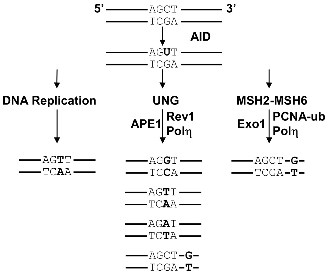
In response to an assault by foreign organisms, peripheral B cells can change their antibody affinity and isotype by somatically mutating their genomic DNA. The ability of a cell to modify its DNA is exceptional in light of the potential consequences of genetic alterations to cause human disease and cancer. Thus, as expected, this mechanism of antibody diversity is tightly regulated and coordinated through one protein, activation-induced deaminase (AID). AID produces diversity by converting cytosine to uracil within the immunoglobulin loci. The deoxyuracil residue is mutagenic when paired with deoxyguanosine, since it mimics thymidine during DNA replication. Additionally, B cells can manipulate the DNA repair pathways so that deoxyuracils are not faithfully repaired. Therefore, an intricate balance exists which is regulated at multiple stages to promote mutation of immunoglobulin genes, while retaining integrity of the rest of the genome. Here we discuss and summarize the current understanding of how AID functions to cause somatic hypermutation.


Figures

FIGURE 1
FIGURE 1
Amino acids defining AID activity. Amino acids have been identified that bind co-factors, cause catalysis, recognize the hotspot motif, and shuttle the protein in and out of the nucleus. Confirmed residues that function in these activities are shown in the upper diagram. Amino acids that are phosphorylated are marked by asterisks and black bars; NES, nuclear export signal. Putative residues that may function are shown in the lower diagram. These include nuclear localization signals (NLS) in the N-terminal region identified by two groups, NLSa (Patenaude et al., 2009), in gray bars, and NLSb (Ito et al., 2004) in black bars. Two residues that may cause retention of AID in the cytoplasm are shown in the C-terminal region (Patenaude et al., 2009).
FIGURE 2
FIGURE 2
Targeting elements for SHM in the Ig loci. For all loci, the relative mutation frequency is shown by shaded peaks over the V region or S region. P, promoter; arrows, start of transcription; iE, intronic enhancer; triangles, location of donor splice sites in the leader exon, V(D)J exon, and iEμ intronic exon. AID may be active in the V regions by transcription bubbles, and in the S regions by R-loops. 3’ enhancers downstream of the C genes are shown on a different scale. For the mouse Igh chain locus, 3’ enhancers include 4 hypersensitive (HS) sites. For the mouse Igκ locus, there are two enhancers, 3’E and Ed. For the chicken DT40 Igλ locus, identified enhancers are 3’E and a regulatory region, 3’RR.
FIGURE 3
FIGURE 3
Three error-prone pathways for processing U:G mismatches. AID deaminates dC to dU in a hotspot, WGCW. Mutations are shown in bold. (1) DNA replication. U mimics T, and replication from the 5’ strand will incorporate C:G to T:A transitions. (2) UNG recognition. UNG removes the U, and several possibilities could occur. Rev1 may bypass the abasic site with a C:G to G:C transversion. APE1 may nick the abasic site, and the 5’ strand would be extended by a low-fidelity polymerase to insert other transversions. Polη may extend the synthesis to insert G opposite a downstream template T, to cause A:T to G:C transitions. (3) MSH2-MSH6 recognition. The two proteins bind to the U:G mismatch and recruit Exo1 to form a gap. It is not known what triggers a nick for Exo1 to access the DNA. Mono-ubiquitinated PCNA brings in Polη to synthesize predominantly A:T mutations downstream of the initial deamination event.

References

    1. Alrefai RH, Winter DB, Bohr VA, Gearhart PJ. Nucleotide excision repair in an immunoglobulin variable gene is less efficient than in a housekeeping gene. Mol Immunol. 2007;44:2800–2805. - PMC - PubMed
    1. Aoufouchi S, Faili A, Zober C, D'Orlando O, Weller S, Weill JC, Reynaud CA. Proteasomal degradation restricts the nuclear lifespan of AID. J Exp Med. 2008;205:1357–1368. - PMC - PubMed
    1. Arakawa H, Hauschild J, Buerstedde JM. Requirement of the activation-induced deaminase (AID) gene for immunoglobulin gene conversion. Science. 2002;295:1301–1306. - PubMed
    1. Arakawa H, Moldovan GL, Saribasak H, Saribasak NN, Jentsch S, Buerstedde JM. A role for PCNA ubiquitination in immunoglobulin hypermutation. PLoS Biol. 2006;4:e366. - PMC - PubMed
    1. Bachl J, Carlson C, Gray-Schopfer V, Dessing M, Olsson C. Increased transcription levels induce higher mutation rates in a hypermutating cell line. J Immunol. 2001;166:5051–5057. - PubMed

Publication types

Substances

LinkOut - more resources