Comparative Study
. 2010 May 27;66(4):523-35.
doi: 10.1016/j.neuron.2010.04.038.

Cux1 and Cux2 regulate dendritic branching, spine morphology, and synapses of the upper layer neurons of the cortex

Affiliations
Comparative Study

Cux1 and Cux2 regulate dendritic branching, spine morphology, and synapses of the upper layer neurons of the cortex

Beatriz Cubelos et al. Neuron. .

Abstract

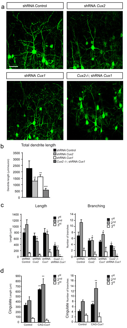
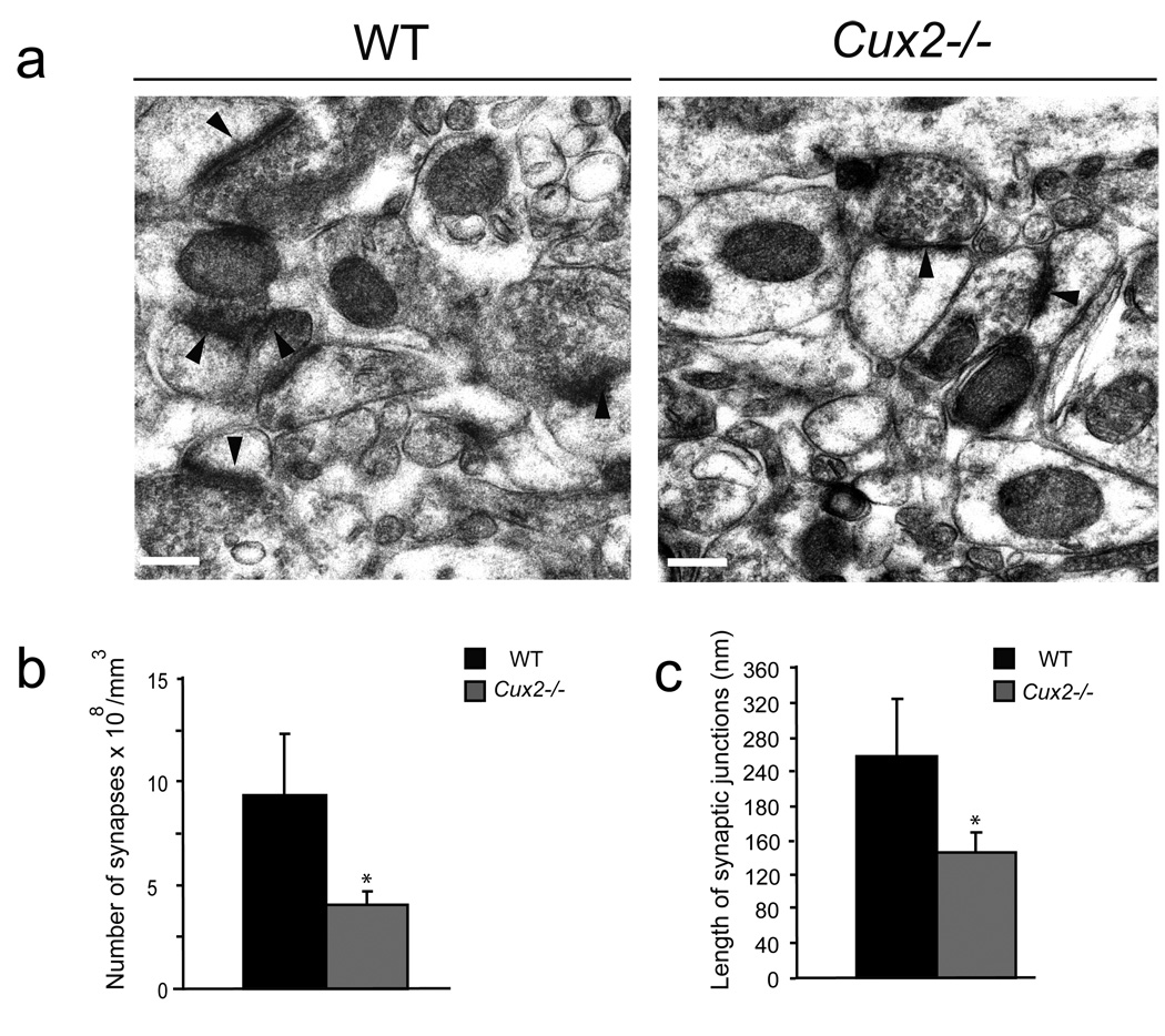
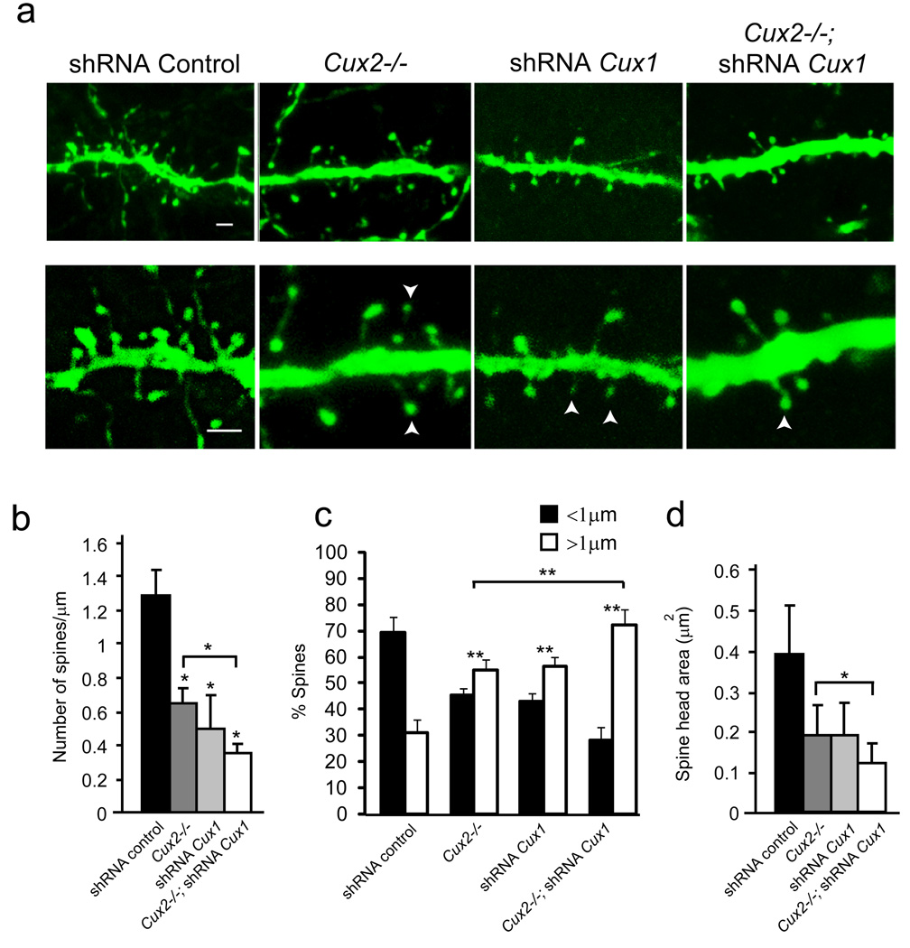
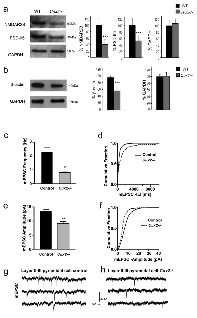
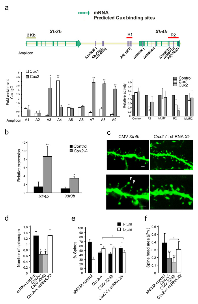
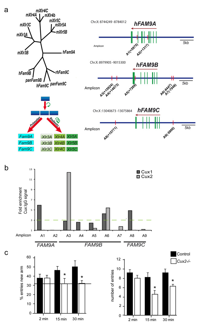
Dendrite branching and spine formation determines the function of morphologically distinct and specialized neuronal subclasses. However, little is known about the programs instructing specific branching patterns in vertebrate neurons and whether such programs influence dendritic spines and synapses. Using knockout and knockdown studies combined with morphological, molecular, and electrophysiological analysis, we show that the homeobox Cux1 and Cux2 are intrinsic and complementary regulators of dendrite branching, spine development, and synapse formation in layer II-III neurons of the cerebral cortex. Cux genes control the number and maturation of dendritic spines partly through direct regulation of the expression of Xlr3b and Xlr4b, chromatin remodeling genes previously implicated in cognitive defects. Accordingly, abnormal dendrites and synapses in Cux2(-/-) mice correlate with reduced synaptic function and defects in working memory. These demonstrate critical roles of Cux in dendritogenesis and highlight subclass-specific mechanisms of synapse regulation that contribute to the establishment of cognitive circuits.


Figures

Figure 1
Figure 1. Cux1 and Cux2 control the dendritic morphology and spine number of upper cortical pyramidal neurons
a, b) Golgi-Cox stained individual neurons in WT, Cux2−/− and Cux1−/− animals. a) Pyramidal neurons in upper cortical layers II–III show fewer dendritic branches and spines in Cux2−/− and Cux1−/− mutants than in the WT animals, (upper panels). High optical magnification images of dendritic spines (lower panels). b) No differences were observed in the dendritic morphology of pyramidal neurons in cortical layer V (upper panels) or in their dendritic spines (lower panels). Bars represent 50 µm (upper panels) and 20 µm (lower panels). c) Total cumulative length of dendritic processes per neuron in cortical layers II–III and V of the somatosensory cortex of WT, Cux1−/− and Cux2−/− mice. d) Dendritic spine density in layers II–III and layer V. e) Total cumulative dendrite length of primary, secondary and tertiary branches per neuron in layers II–III. f) Total number of primary, secondary and tertiary dendrite branches per neuron in layers II–III. WT (n= 16), Cux1−/− (n=15) and Cux2−/− (n=15). * p<0.05 and ** p<0.01 between WT and mutant cortex.
Figure 2
Figure 2. Cux1 and Cux2 proteins stimulate dendrite development via cell intrinsic and additive mechanisms
a) Confocal micrographs showing GFP-expressing layer II–III neurons in the P21 cortex. Neuronal morphology was analyzed at P21 after in utero electroporation at E15.5. Knock-down of Cux1 or Cux2 with shRNA lentiviral constructs decreases the dendrite complexity of layer II–III neurons compared to control shRNA electroporated neurons. Knock-down of Cux1 in Cux2−/− layer II–III neurons induces still simpler dendrite morphologies. Bar represents 25 µm. b) Total cumulative lengths of dendritic processes per GFP-positive neuron in layers II–III. c) Cumulative dendrite length of primary, secondary and tertiary branches (left) and the average number of primary, secondary and tertiary dendrite branches (right) per neuron. Control shRNA (n= 19), shRNA Cux1 (n=15) and shRNA Cux2 (n=22), shRNA Cux1 in Cux2−/− (n=12). d) Overexpression of Cux1 in neurons of the cingulate cortex stimulates dendritic branching. Cumulative dendrite length of primary, secondary and tertiary branches (left) and the number of primary, secondary and tertiary dendrite branches (right) per GFP positive layer II–III neuron control (n= 15), CAG Cux1 (n=15) * p<0.05, ** p<0.01 and *** p<0.001 compared with controls.
Figure 3
Figure 3. Altered synapse formation in the upper layers of Cux2−/− mice
a)Electron micrographs showing the synapses (arrowheads) in sections of cortical layers II–III of the somatosensory cortex of WT and Cux2−/− animals. Bar represents 0.25 µm. b) Quantification of synapse density in layers II–III of WT and Cux2−/− animals. c) Average length of the synaptic junction apposition surface in layers II–III of WT and Cux2−/− animals. * p<0.001 compared with WT.
Figure 4
Figure 4. Cux1 and Cux2 regulate dendritic spine number and spine morphology
a) Confocal images, showing dendritic spines of GFP-positive layer II–III neurons expressing control, Cux1, or Cux2 shRNAs and of WT or Cux2−/− P21 cortex. Bar represents 1 µm. Arrowheads point to small spine heads. b, c, d) Quantitative analysis of dendritic spine defects. n≥ 15 dendrite segments and n≥ 500 spines for each sample. * p<0.01 and ** p<0.001 compared to WT or Cux2−/− (brackets).
Figure 5
Figure 5. Reduced expression of synaptic proteins and changes in layer II–III mEPSC in amplitude and frequency in Cux2−/−
a, b) Reduced expression of synaptic proteins in Cux2−/−.Western blot analysis of the expression of NMDAR2B, PSD-95 (a) and β-actin (b) in total cortical lysates from WT (n=4) and Cux2−/− (n=4). Graphs show the mean and SD signal quantification of the relative amount of protein in WT and Cux2−/− cortices. *** p<0.001. c) Average frequency of mEPSC of layer II–III pyramidal cells from control (WT and Cux2+/−) and Cux2−/− mice. (* p< 0.0005, Student’s, unpaired t test, n=13 and 14 cells, respectively), d) Cumulative fraction curves of interevent intervals (IEIs) for mEPSC of layer II–III pyramidal cells showing longer IEIs in Cux2−/− compared with control (p < 0.0005, K–S test). e) Average amplitude of mEPSC in layer II–III pyramidal cells from Cux2−/− (** p < 0.0005, Student’s, unpaired t test, n=13 and 14 cells, respectively). f) Cumulative fraction curves of amplitude of layer II–III pyramidal cells showing smaller amplitude in Cux2−/− animals compared with control (p < 0.0005, K-S test). g, h) Representative traces of mEPSC from layer II–III pyramidal cells of control and Cux2−/− mice, respectively. Data in bar graphs depict mean + SEM; control: black bars; Cux2−/−: gray bars. IEI: Interevent interval. mEPSC: miniature excitatory postsynaptic current.
Figure 6
Figure 6. Cux1 and Cux2 regulate dendritic spine number and spine morphology through mechanisms that involve the repression of Xlr genes
a) Cux putative binding sites identified (MatInspector (Genomatrix)) in the genomic region containing the Xlr gene cluster (see graphic). Left diagram, in vivo chromatin immunoprecipitation. 400bp average chromatin fragments were obtained from adult cortex and immunoprecipitation with Cux1 and Cux2 antibodies was performed. Binding to nine regions was tested by Q-PCR. Relative positions of the amplicons (A) to the Xlr4b ATG (+1) are indicated. Real Time PCR reactions were carried out in duplicates in three independent preparations of immunoprecipitated material from three cortexes. The fold enrichment for each tested region was normalized to control IgG.*p<0.01 and **p<0.001 compared to control IgG or region 1. Right graph, luciferase experiments performed in neuronal cells obtained from E12 cortex. Cux1 and Cux2 repress transcriptional activity of luciferase construct reporters containing regions R1 and R2 but not of these reporters when Cux putative sites are mutated (mutR1 and mutR2). *p<0.01 and **p<0.001 b) Up-regulation of Xlr4b and Xlr3b in the adult Cux2−/− cortex. Relative expression of Xlr4b and Xlr3b mRNA is shown in relation to one control sample normalized as 1. Expression of Xlr genes is shown as the ratio of the amounts of Xlr and GADPH transcripts measured by Q-PCR in total RNA obtained from the cortex of adult male Cux2+/− (n=4) and Cux2−/− (n=4) animals. * p<0.2 and ** p<0.05. c) Reduced number and aberrant morphologies of dendritic spines in GFP-positive layer II–III neurons over-expressing Xlr4b in WT animals (left panels). Reverted dendritic spine phenotypes in layer II–III neurons of Cux2−/− electroporated with shRNAs targeting Xlr genes (right panels). Bar represents 1 µm. Arrowheads point to small spine heads. d, e, f) Quantitative analysis of dendritic spine defects in GFP-positive layer II–III neurons with the indicated shRNAs. n 15 dendrite segments and n≥ 500 spines for each sample.* p<0.01 and ** p<0.001 compared to WT or Cux2−/− (brackets).
Figure 7
Figure 7. Human FAM9 genes and cognitive defects
a) Left diagram shows the phylogenetic relationship between Xlr and FAM9 superfamily members. Below, the possible duplication of an ancestral gene that gave rise to the Xlr and FAM9 orthologous genes. The upper right panel schematizes the location of putative Cux binding sites in FAM9A, B and C genes. b) Immunoprecipitation of the putative binding sites with anti-Cux1 and anti-Cux2 was tested in BE(2)-M17 human neuroblastoma cells transfected with Cux1 or Cux2 and by semi-quantitative PCR (representative experiment of three independent experiments). Relative positions of the amplicons (A) to each ATG (+1) are indicated. c) Cux2−/− mice have defects in working memory. Working memory was assessed in control and Cux2−/− mice with a two–trial memory task based on free-choice exploration of a Y-maze. ITI: inter-trial intervals (see Experimental Procedures). Histograms show the percentage of visits (left panel) and number of total visits to the new arm (right panel). Control and Cux-2−/− animals showed no differences in exploratory behavior (ITI=2 min), but working memory was impaired in Cux-2−/− mice (ITIs of 15 and 30 min).
Figure 8
Figure 8. Cux1 and Cux2 promote dendritic branching and spine differentiation
Cux1 and Cux2 induce cell autonomous development of dendritic branches and promote dendritic spine development and stabilization in early differentiating neurons by at least partly independent mechanisms. Regulation of Xlr3b and Xlr4b gene expression by Cux proteins contributes to trigger dendritic spine differentiation.

References

    1. Ammer AG, Weed SA. Cortactin branches out: roles in regulating protrusive actin dynamics. Cell motility and the cytoskeleton. 2008;65:687–707. - PMC - PubMed
    1. Arion D, Unger T, Lewis DA, Mirnics K. Molecular markers distinguishing supragranular and infragranular layers in the human prefrontal cortex. The European journal of neuroscience. 2007;25:1843–1854. - PubMed
    1. Ballesteros-Yanez I, Benavides-Piccione R, Elston GN, Yuste R, DeFelipe J. Density and morphology of dendritic spines in mouse neocortex. Neuroscience. 2006;138:403–409. - PubMed
    1. Benavides-Piccione R, Hamzei-Sichani F, Ballesteros-Yanez I, DeFelipe J, Yuste R. Dendritic size of pyramidal neurons differs among mouse cortical regions. Cereb Cortex. 2006;16:990–1001. - PubMed
    1. Bourne J, Harris KM. Do thin spines learn to be mushroom spines that remember? Current opinion in neurobiology. 2007;17:381–386. - PubMed

Publication types

Associated data