. 2010 May 28;141(5):775-85.
doi: 10.1016/j.cell.2010.03.055.

Hematopoietic origin of pathological grooming in Hoxb8 mutant mice

Affiliations

Hematopoietic origin of pathological grooming in Hoxb8 mutant mice

Shau-Kwaun Chen et al. Cell. .

Abstract

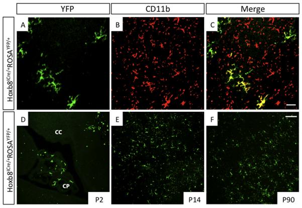
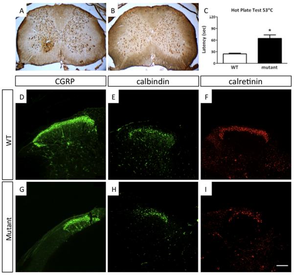
Mouse Hoxb8 mutants show unexpected behavior manifested by compulsive grooming and hair removal, similar to behavior in humans with the obsessive-compulsive disorder spectrum disorder trichotillomania. As Hox gene disruption often has pleiotropic effects, the root cause of this behavioral deficit was unclear. Here we report that, in the brain, Hoxb8 cell lineage exclusively labels bone marrow-derived microglia. Furthermore, transplantation of wild-type bone marrow into Hoxb8 mutant mice rescues their pathological phenotype. It has been suggested that the grooming dysfunction results from a nociceptive defect, also exhibited by Hoxb8 mutant mice. However, bone marrow transplant experiments and cell type-specific disruption of Hoxb8 reveal that these two phenotypes are separable, with the grooming phenotype derived from the hematopoietic lineage and the sensory defect derived from the spinal cord cells. Immunological dysfunctions have been associated with neuropsychiatric disorders, but the causative relationships are unclear. In this mouse, a distinct compulsive behavioral disorder is associated with mutant microglia.


Figures

Figure 1
Figure 1. Hoxb8 Cell Lineage Gives Rise to Brain Microglia
(A-F) Analysis of Hoxb8 lineage in mice heterozygous for the Hoxb8-IRES-Cre and ROSA-YFP alleles. To determine if cells of Hoxb8 lineage in the brain are microglia, the identity of YFP-positive cells was examined by immunohistochemistry. Sagittal sections of the adult cerebral cortex were co-stained with (A) anti-GFP antibody and (B) anti-CD11b antibody. (C) Co-localization of both signals shows that these cells are microglia. (D) Cortical microglia originating from the Hoxb8 cell lineage first appear in the brain during the first two postnatal days (P2), in the choroid plexus and in association with the ventricular lining. (E) The number of YFP-positive cells markedly increases by P14 throughout the cerebral cortex. This high abundance is maintained in the adult life (F). CP, Choroid plexus; CC, cerebral cortex. See supplemental Figure 1.
Figure 2
Figure 2. Hoxb8 Cell Lineage Labels All Hematopoietic Groups Examined
(A-F) Peripheral white blood cells from Hoxb8-IRES-Cre; ROSA-YFP double heterozygotes were collected and the YFP signal examined by FACS. (A-C) Control blood samples from ROSA-YFP reporter mice in the absence of the Hoxb8-IRES-Cre driver. (D-F) Analysis of blood samples collected from ROSA-YFP reporter mice combined with the Hoxb8-ICre driver. Markers used: (A and D) Mac1/ Gr-1 and YFP; (B and E) CD19 and YFP; (C and F) CD4/ CD8 and YFP. (G) YFP signal was detected by fluorescence microscopy in the majority of bone marrow cells. (H) Most of the cells in the hematopoietic stem cell and multipotent progenitor cell domain are YFP positive. Left panel: FACS analysis with Sca-1 and c-kit markers. The cells shown in the rectangle were further analyzed for YFP fluorescence. Top right panel: the black histogram represents YFP fluorescence detected in cells collected from ROSA-YFP reporter mice in the absence of the Hoxb8-ICre driver, while the white histogram (bottom right panel) represents cells collected from ROSA-YFP reporter mice carrying the Hoxb8-IRES-Cre driver. See supplemental Figure 2.
Figure 3
Figure 3. Rescue of Excessive Grooming and Hair Removal Defect in Hoxb8 Mutant Mice Transplanted with Normal Bone Marrow
(A) Hoxb8 mutant transplanted with normal bone marrow showing typical hair loss four weeks after transplantation. (B) Hoxb8 mutant mouse three months after transplantation with wild-type bone marrow cells showing complete recovery from hair loss. (C) A close-up view of the ventral anterior part of the body, which is the primary region of hair removal. (D) Laboras data collected over a 24-hour period with Hoxb8 mutant mice transplanted with wild-type bone marrow cells, show significant decrease in grooming times relative to Hoxb8 mutant mice. White bar represents wild-type controls (n=22) relative to Hoxb8 mutants (n=25). Grey bar indicates the grooming time of Hoxb8 mutant mice rescued by normal bone marrow transplants (n=6) *p<0.05 versus mutant. (E) A wild-type mouse, transplanted with Hoxb8 mutant bone marrow, showing a hair removal and lesion pattern typical of Hoxb8 mutant mice. (F) Grooming times of two wild-type mice transplanted with mutant bone marrow that developed hairless patches. These experimental animals (grey column, n=2) showed elevated grooming times, although not as long as the average observed in a large cohort of Hoxb8 mutants *p<0.05 versus wild-type. See supplemental Figure 3. Columns represent the mean ± 1SEM.
Figure 4
Figure 4. Anatomical and Nociceptive Defects in Hoxb8 Mutant Mice
(A and B) Spinal cord sections at cervical levels in (A) wild-type mice and (B) Hoxb8 mutants stained with anti-NeuN antibody. These sections are representative of wild-type and Hoxb8 mutant sections taken along the spinal cord from C4 through the lumbar region L5. Neuron counts are decreased and the remaining interneurons noticeably disorganized in the mutant spinal laminae. (C) The latency of response to heat at 53°C, was significantly increased in Hoxb8 mutants. White bar, control siblings; black bar, Hoxb8 mutant mice. Columns represent the mean ± 1SEM. (D - K) Anatomical defects in dorsal spinal cord of Hoxb8 mutant mice. Spinal cord sections shown at L4-L5 from (D-F) wild-type mice and (G-I) Hoxb8 mutant mice. The spinal cord sections were labeled with a marker for nociceptive sensory fibers (CGRP), and with interneuron markers for lamina I and II (calbindin and calretinin). The number of interneurons in laminae I and II, are decreased and disorganized in Hoxb8 mutant mice relative to wild-type mice. Scale bar (D – I). 100μM. See supplemental Figure 4.
Figure 5
Figure 5. Nociceptive Defects in Hoxb8 Mutant Mice Were Not Rescued by Transplantations with Wild-Type Bone Marrow
Hoxb8 mutant mice, whose pathological grooming defects were rescued by normal bone marrow transplants, still exhibit significantly longer latency of response to heat (53°C Hot Plate Test), comparable to the Hoxb8 mutant mice. White column, wild-type controls (n=14); black column, Hoxb8 mutants (n=11), grey column, Hoxb8 mutants phenotype-rescued with normal bone marrow (n=6). Data were collected 4-5 months after bone marrow transplantation. Columns represent the mean ± 1SEM. *p<0.05 versus wild-type.
Figure 6
Figure 6. Mice with Hoxb8 Deletion Restricted to the Hematopoietic System Develop Typical Excessive Grooming and Hair Removal Phenotype but not Nociceptive Spinal Cord Defects
(A) Tie2 lineage is present in CNS microglia. Brains and spinal cords from mice carrying the Tie2Cre transgene and ROSA-YFP allele were stained with anti-GFP antibodies. The microglial identity was confirmed by double staining with CD11b (not shown). (B) Conditional inactivation of the Hoxb8 locus restricted to the hematopoietic system recapitulates hair removal and excessive grooming phenotype: Hairless patches developing in the shoulder and chest area of a 3-month-old conditional mutant mouse. (C) Conditional mutant mice (n=3) exhibit excessive grooming compared to control siblings (n=5). (D) Tie2Cre conditional mutant mice (black column) do not show increased latency times in response to heat relative to control mice (white bar). (E-J) No histological defects analogous to those in Hoxb8 mutant mice were found in dorsal spinal laminae of Tie2Cre conditional mutants. Spinal cord sections at L4-L5 levels were collected from wild-type mice, Hoxb8 mutant mice and Tie2Cre conditional Hoxb8 mutants. Interneurons in laminae I and II were stained for calbindin (E-G) and calretinin (H-J). The regions with neurons positively staining for these markers are circumscribed with white dashed boundary. Scale Bar: (A), 50 μM, (E-J), 100 μM. Columns represent the mean ± 1SEM. *p<0.05.
Figure 7
Figure 7. Conditional Deletion of Hoxb8 in the Hoxc8 Domain Recapitulates Nociceptive Defects But Not Excessive Grooming or Hair Removal Behavior
(A) Hoxc8 lineage is present in all laminae of the spinal cord. X-gal staining was performed in spinal cord sections collected from mice carrying both Hoxc8-IRES-Cre and ROSA-LacZ alleles. (B) Hoxc8 cell lineage in brain. Brain sections collected from Hoxc8-IRES-Cre; ROSA-YFP mice were stained with anti-GFP antibody. (C). Compared to the Hoxb8 lineage, only a small percentage (<3%) of ramified microglia were labeled by the Hoxc8 lineage. The percentage of YFP positive cells to total ramified microglia (Iba1 positive cells) was determined from sections through cerebral cortex derived from three Hoxb8-IRES-Cre/Rosa-YFP (black column) and three Hoxc8-IRES-Cre/Rosa26-YFP mice (white column). (D) No hair removal and excessive grooming were detected in Hoxc8-IRES-Cre conditional mutants (10/10 animals). (E) No significant difference in grooming time between four month old Hoxc8-IRES-Cre-conditional Hoxb8 mutants and control siblings were observed. (n=4). (F) Hoxc8-Cre conditional mutants exhibit heat insensitivity very similar to that observed in Hoxb8 mutant mice. (G-L) The numbers of interneurons in laminae I and II are decreased in Hoxc8Cre-conditional Hoxb8 mutants. Spinal cord sections at L4-L5 levels were collected from (G and J) wild-type mice, (H and K) Hoxb8 mutant mice, and (I and L) Hoxc8Cre-conditional Hoxb8 mutants, and stained for calbindin (G-I) and calretinin (J-L). The positive regions are highlighted with white dashed line. Scale Bar: (B), (G-L), 100 μM. Columns represent the mean ± 1SEM. *p<0.05.

Comment in

References

    1. Aldridge JW, Berridge KC, Herman M, Zimmer L. Neuronal coding of serial order: syntax of grooming in the neostriatum. Psychol Sci. 1993;4:391–395.
    1. Alliot F, Godin I, Pessac B. Microglia derive from progenitors, originating from the yolk sac, and which proliferate in the brain. Brain Res Dev Brain Res. 1999;117:145–152. - PubMed
    1. Ashwood P, Wills S, Van de Water J. The immune response in autism: a new frontier for autism research. J Leukoc Biol. 2006;80:1–15. - PubMed
    1. Berridge KC. Progressive degradation of serial grooming chains by descending decerebration. Behav Brain Res. 1989;33:241–253. - PubMed
    1. Berridge KC, Fentress JC, Parr H. Natural syntax rules control action sequence of rats. Behav Brain Res. 1987;23:59–68. - PubMed

Publication types