. 2010 May 28;141(5):834-45.
doi: 10.1016/j.cell.2010.03.052.

A bivalent tarantula toxin activates the capsaicin receptor, TRPV1, by targeting the outer pore domain

Affiliations

A bivalent tarantula toxin activates the capsaicin receptor, TRPV1, by targeting the outer pore domain

Christopher J Bohlen et al. Cell. .

Abstract

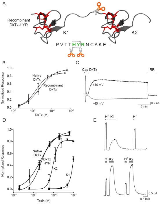
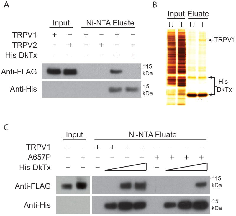
Toxins have evolved to target regions of membrane ion channels that underlie ligand binding, gating, or ion permeation, and have thus served as invaluable tools for probing channel structure and function. Here, we describe a peptide toxin from the Earth Tiger tarantula that selectively and irreversibly activates the capsaicin- and heat-sensitive channel, TRPV1. This high-avidity interaction derives from a unique tandem repeat structure of the toxin that endows it with an antibody-like bivalency. The "double-knot" toxin traps TRPV1 in the open state by interacting with residues in the presumptive pore-forming region of the channel, highlighting the importance of conformational changes in the outer pore region of TRP channels during activation.


Figures

Figure 1
Figure 1. The Chinese bird spider produces a novel bivalent TRPV1 toxin
(A) The Chinese bird spider (Ornithoctonus huwena) is a large terrestrial tarantula with a leg span of up to 12 cm. It is found primarily in the Guangxi province of China (photo courtesy of Chuck Kristensen, SpiderPharm, Inc.). (B) Purified DkTx toxin evokes robust calcium increases in HEK293 cells expressing the rat TRPV1 channel. Co-application of ruthenium red (RR; 10 μM), a non-selective TRP channel pore blocker, inhibits toxin-evoked responses. Purple denotes low resting cytoplasmic calcium and orange indicates calcium increase. (C) DkTx is a new member of the ICK peptide family. Other than the highly conserved arrangement of cysteine residues (highlighted in yellow), DkTx shows no obvious sequence similarity with other ICK peptides, including the vanillotoxins (VaTx1-3) and hanatoxin (HaTx). (D) DkTx consists of two ICK lobes (Knot 1 and Knot 2) separated by a short linker region. The NMR solution structure of HaTx (Takahashi et al., 2000) served as a template for a hypothetical model of DkTx, showing the conserved disulfides in yellow for HaTx and red for DkTx (generated using PyMOL, http://www.pymol.org). These tandemly repeated lobes show significant sequence identity (see panel C), suggesting that they arose by gene duplication.
Figure 2
Figure 2. DkTx is a selective and irreversible TRPV1 activator
(A) Trigeminal sensory neurons from wild type (V1+/+) or TRPV1-deficient (V1-/-) mice were examined for responses to capsaicin (1 μM; Cap) and DkTx (5 μM) using ratiometric calcium imaging. Depolarization with high extracellular potassium (75 mM; Hi K) identified all neurons in the field. (Left) Pseudocolor images of Fura-2-loaded cells (color bar indicates relative change in fluorescence ratio, with purple and white denoting lowest and highest cytoplasmic calcium). (Right) Average ratiometric calcium responses as a function of time. Solid and dashed lines represent responses from wild type and TRPV1-deficient trigeminal neurons, respectively. Note lack of DkTx-evoked response by TRPV1-deficient neurons (n ≥ 50 cells per trace). (B) Both capsaicin (Cap; 1 μM) and DkTx (2 μM) elicited outwardly rectifying, ruthenium red (RR; 4 μM) blockable currents in cultured mouse trigeminal neurons (recorded in whole-cell patch clamp configuration). Current-voltage relationships and representative current trace are shown at left and right, respectively. Note persistence of DkTx-evoked response even after 3-4 min washout period. (C) (Left) Relative washout rates were determined for electrophysiological responses to saturating doses of capsaicin (1 μM; orange), VaTx2 (2 μM; purple), or DkTx (2 μM; green). Individual points represent fractional current remaining as measured by whole-cell patch-clamp recording from TRPV1-expressing HEK293 cells (Vh = +80 mV). When washout was complete, a time constant of decay could be determined by exponential fit of the data (τoff = 0.17 min and 1.3 min for capsaicin and VaTx2, respectively) (n = 4-7 cells per point). (Right) Plot of fractional DkTx-evoked current remaining after a 1, 5, or 15 min washout period as determined for two different toxin concentrations (n = 5-9 cells per bar). Average values represent mean ± s.e.m.
Figure 3
Figure 3. Bivalency is required for persistent toxin action
(A) Schematic depicts location of engineered protease (Genenase I) cleavage site in recombinant DkTx. (B) Native and recombinant DkTx are equipotent as determined by calcium imaging using TRPV1-expressing HEK293 cells. Values are normalized to maximal capsaicin (10 μM)-evoked responses (n = 3-4 wells per point). (C) Whole cell patch-clamp recording from TRPV1-expressing HEK293 cells shows that recombinant DkTx (2 μM) produces persistent, ruthenium red (RR) blockable membrane currents comparable in magnitude to those elicited by capsaicin (1 μM). (D) Dose-response analysis (carried out as in ‘B’) shows that native and recombinant DkTx containing the Genenase I cleavage site (HYR) are equipotent. Knot 1 and Knot 2 peptides (K1 and K2), derived from Genenase I cleavage of HYR, are active but significantly less potent (EC50 = 8.9 and 0.97 μM respectively). Values are normalized to maximal capsaicin (10 μM)-evoked responses (n = 3-4 wells per point). (E) Representative whole cell patch-clamp recordings (+80 mV) from TRPV1-expressing HEK293 cells showing responses to extracellular protons (H+; pH 5.5), K1 (20 μM; top trace) or K2 (20 μM; bottom trace) peptides. Note rapidly reversible (i.e. non-persistent) nature of K1- or K2-evoked responses following washout. Average values represent mean ± s.e.m.
Figure 4
Figure 4. The TRPV1 pore domain specifies toxin sensitivity
(A) Chimeric channels were generated between rat and Xenopus TRPV1 orthologues (red and blue bars, respectively) based on the location of putative transmembrane domains (grey bars) All chimeras shown were activated by capsaicin. Selective sensitivity of rat TRPV1 to DkTx mapped to residue A657, which when replaced by the equivalent residue from the frog channel (P663) led to dramatically reduced toxin sensitivity. (B) Representative whole cell patch clamp recording (+80 mV) from HEK293 cells expressing the rat TRPV1 (A657P) mutant showed selective loss of DkTx (2 μM; left) or VaTx3 (2 μM; center) sensitivity. In contrast, sensitivity to capsaicin (Cap; 1 μM) and extracellular protons (H+; pH 5.5) was retained. Co-application of capsaicin and DkTx (right) failed to produce persistent channel activation. (C) Representative two-electrode voltage clamp recordings (+80 mV) from Xenopus oocytes expressing wild type frog TRPV1 channel (left) shows specific insensitivity to DkTx (20 μM) versus capsaicin and protons (50 μM at pH 5.5). The frog TRPV1 P663A mutant (center) showed acquisition of toxin sensitivity. Note reversibility of toxin-evoked current. Graph at right shows average toxin responses for wild type rat, wild type frog, and mutant frog (P663A) channels normalized to capsaicin-evoked responses (n = 3-5 cells per bar; average values represent mean ± s.e.m.).
Figure 5
Figure 5. DkTx binds directly to TRPV1 via association with the pore domain
(A) DkTx affinity resin retains detergent solubilized TRPV1, but not TRPV2 protein. Recombinant His-tagged DkTx was immobilized on nickel affinity (Ni-NTA) resin, to which affinity purified FLAG-tagged TRPV1 or TRPV2 protein (Input) was subsequently applied. After extensive washing, toxin-channel complexes were eluted and analyzed by Western blotting using anti-FLAG or anti-His antisera to detect channel or toxin protein, respectively. Complexes were observed with TRPV1, but not TRPV2 input, and were not detected with Ni-NTA beads lacking toxin. (B) Crude membrane extracts (Input) from uninduced (U) or induced (I) TRPV1-HEK293 cells were applied to DkTx-coupled resin. Bound material (Eluate) was recovered and analyzed by silver staining, showing substantial enrichment of TRPV1-toxin complexes over other membrane proteins. (C) TRPV1 A657P mutant binds less avidly to DkTx affinity resin. Purified wild type (TRPV1) and mutant (A657P) protein were analyzed for toxin binding as described in (A) using resins with increasing DkTx substitution (wedges depict 3-fold concentration range). Mutant (A657P) channel protein was retained only at the highest toxin density.
Figure 6
Figure 6. Mutagenesis outlines a DkTx footprint on the extracellular face of TRPV1
(A) Representative voltage-clamp recordings (Vh = +80 mV) from oocytes expressing wild type or mutant rat TRPV1 channels in response to double knot toxin (DkTx; 1.5 μM), then capsaicin (Cap; 3 μM), followed by block with ruthenium red (RR; 80 mM). (B) Quantitative comparison of toxin-evoked responses normalized to a maximal capsaicin-evoked response. Note substantial and significant (n = 5 – 8 cells per mutant; average values represent mean ± s.e.m.; p < 0.01, one-way ANOVA) decrement in toxin sensitivity for all mutants shown versus wild type TRPV1. (C) Whole cell patch-clamp recording from a transfected HEK293 cell expressing a TRPV1 channel containing all four outer pore mutations shows complete loss of toxin sensitivity even when challenged with DkTx at a concentration (20 μM) exceeding the EC50 by 100-fold. (D) Putative locations of residues required for DkTx sensitivity are mapped onto a model pore domain based on the structure of the bacterial potassium channel, KcsA (Doyle et al., 1998). Amino acid side chains of TRPV1 residues I599, F649, A657, and F659 are shown in color, depicting a potential footprint of toxin binding. Red and orange groups correspond to bivalent DkTx attachment sites on adjacent channel subunits. Such an interaction may also be mediated through interaction with non-adjacent (orthogonal) subunits.
Figure 7
Figure 7. VaTx1 targets distinct regions of TRPV1 and Kv2.1
(A) Representative whole cell patch clamp recording (+80 mV) from HEK293 cells expressing rat TRPV1 (top) or the TRPV1 (A657P) mutant (bottom) show VaTx1 (100 μM) activation of the wild type, but not the mutant channel. Capsaicin (Cap; 1 μM) and extracellular protons (H+; pH 5.5) are shown for reference. (B) Two-electrode voltage clamp recordings from Xenopus oocytes expressing rat Kv2.1 voltage-gated potassium channels in the absence (black) and presence (green) of the vanillotoxin VaTx1 (20 μM). Two mutations (F274R and E277K) that are known to abrogate the inhibitory effect of hanatoxin on this channel also eliminated VaTx1-evoked inhibition, whereas a third (I273Y) had no substantial effect on VaTx1 action (n = 3 - 5 cells per trace). (C) Model of the transmembrane topology of TRPV1 (gray bars represent transmembrane helices) highlighting residues (red dots) that are crucial for double-knot toxin (DkTx) activation. In the simplest scenario, the two knots of DkTx bind to two equivalent sites on multiple subunits of the same channel. Kv channels likely possess the same overall transmembrane topology as TRPV1, but interact with ICK toxins in different ways. For example, charybdotoxin (CTx) binds within the ion permeation path to block ion flux, and voltage-modulator toxins, such as hanatoxin (HaTx), target the voltage sensor to modify gating properties (blue and green dots represent mutations that attenuate CTx and HaTx inhibition, respectively). Our findings suggest that single-knot vanillotoxins (VaTx) also target the S3-S4 helices of Kv channels, but activate TRPV1 through the pore region.

References

    1. Alabi AA, Bahamonde MI, Jung HJ, Kim JI, Swartz KJ. Portability of paddle motif function and pharmacology in voltage sensors. Nature. 2007;450:370–375. - PMC - PubMed
    1. Brauchi S, Orio P, Latorre R. Clues to understanding cold sensation: thermodynamics and electrophysiological analysis of the cold receptor TRPM8. Proc Natl Acad Sci U S A. 2004;101:15494–15499. - PMC - PubMed
    1. Bulaj G, Olivera BM. Folding of conotoxins: formation of the native disulfide bridges during chemical synthesis and biosynthesis of Conus peptides. Antioxid Redox Signal. 2008;10:141–155. - PubMed
    1. Catterall WA, Cestele S, Yarov-Yarovoy V, Yu FH, Konoki K, Scheuer T. Voltage-gated ion channels and gating modifier toxins. Toxicon. 2007;49:124–141. - PubMed
    1. Cestele S, Qu Y, Rogers JC, Rochat H, Scheuer T, Catterall WA. Voltage sensor-trapping: enhanced activation of sodium channels by beta-scorpion toxin bound to the S3-S4 loop in domain II. Neuron. 1998;21:919–931. - PubMed

Publication types