. 2010 May 28;141(5):846-58.
doi: 10.1016/j.cell.2010.04.011.

Two cyclin-dependent kinase pathways are essential for polarized trafficking of presynaptic components

Affiliations

Two cyclin-dependent kinase pathways are essential for polarized trafficking of presynaptic components

Chan-Yen Ou et al. Cell. .

Abstract

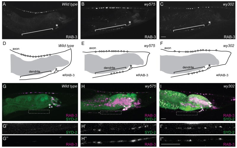
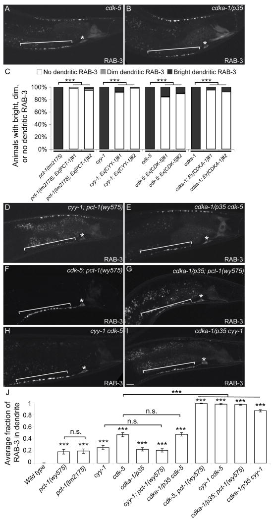
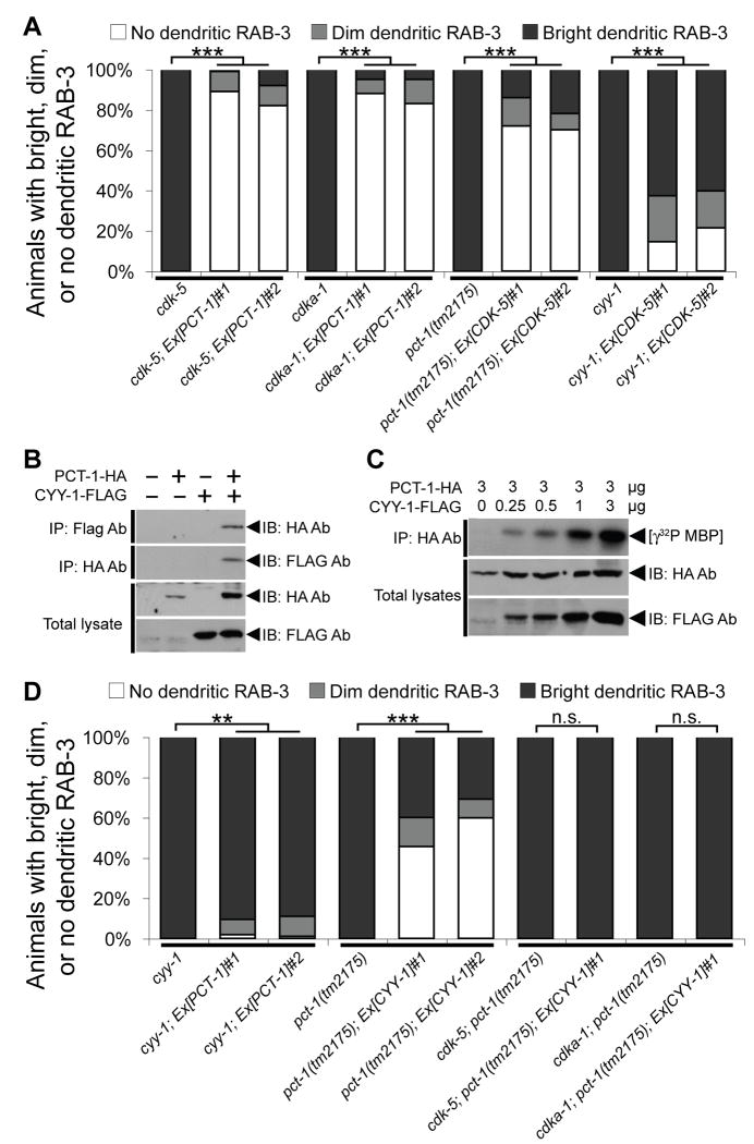
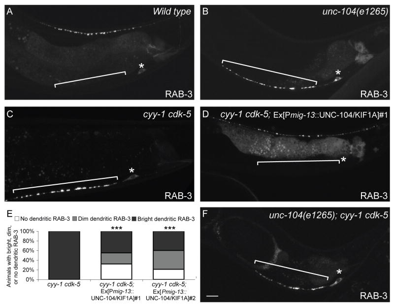
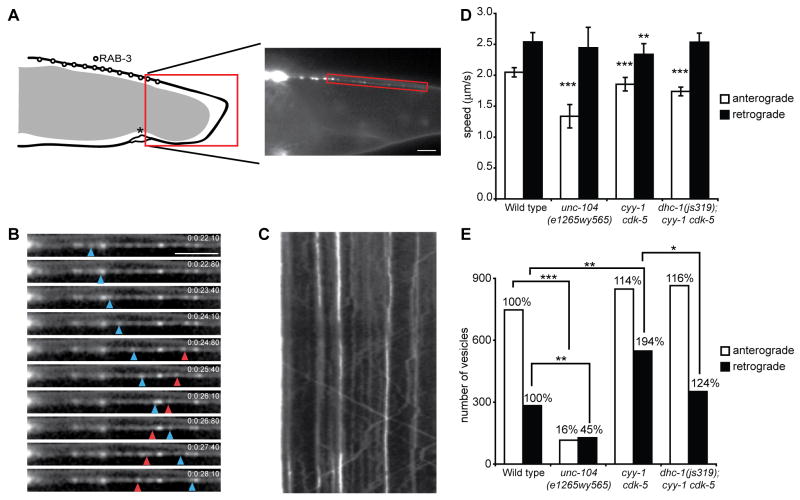
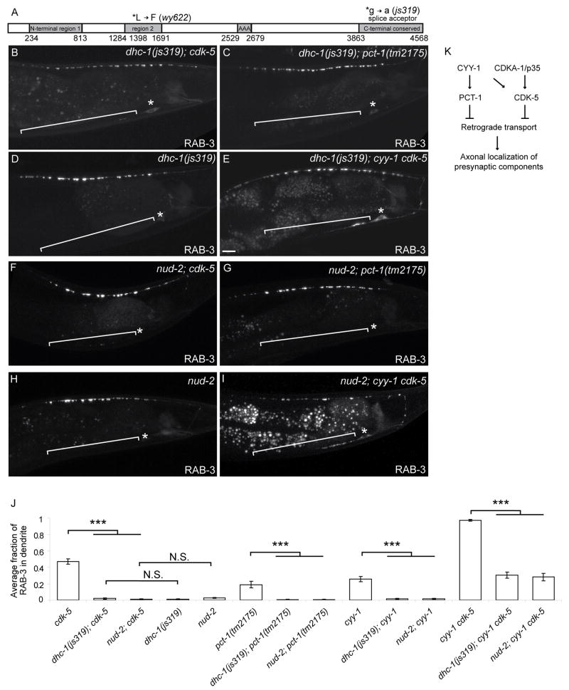
Polarized trafficking of synaptic proteins to axons and dendrites is crucial to neuronal function. Through forward genetic analysis in C. elegans, we identified a cyclin (CYY-1) and a cyclin-dependent Pctaire kinase (PCT-1) necessary for targeting presynaptic components to the axon. Another cyclin-dependent kinase, CDK-5, and its activator p35, act in parallel to and partially redundantly with the CYY-1/PCT-1 pathway. Synaptic vesicles and active zone proteins mostly mislocalize to dendrites in animals defective for both PCT-1 and CDK-5 pathways. Unlike the kinesin-3 motor, unc-104/Kif1a mutant, cyy-1 cdk-5 double mutants have no reduction in anterogradely moving synaptic vesicle precursors (SVPs) as observed by dynamic imaging. Instead, the number of retrogradely moving SVPs is dramatically increased. Furthermore, this mislocalization defect is suppressed by disrupting the retrograde motor, the cytoplasmic dynein complex. Thus, PCT-1 and CDK-5 pathways direct polarized trafficking of presynaptic components by inhibiting dynein-mediated retrograde transport and setting the balance between anterograde and retrograde motors.


Figures

Figure 1
Figure 1
Synaptic Vesicle-Associated RAB-3 and Active Zone Protein SYD-2/Liprin-α Mislocalize to the DA9 Dendrite in wy575 or wy302 Mutants,. (A) A wild-type adult expressing GFP∷RAB-3 in DA9(wyIs85). (B-C) Two mutants expressing GFP∷RAB-3. (D-F) Schematic diagrams of GFP∷RAB-3 distribution. (G-I) Wild-type (G) and mutant animals (H, I) co-expressing GFP∷SYD-2/liprin-α and mcherry∷RAB-3(wyEx2055). (G’-I’, G”-I”) Higher magnification micrographs of the corresponding dotted boxes. The fluorescence in the middle of the worm is gut autofluorescence. Anterior, left and dorsal, top. Bracket, dendrite. Asterisk above cell body. Scale bar, 10μm. See also Figure S1.
Figure 2
Figure 2
PCT-1, CYY-1, CDK-5, and CDKA-1/p35 Act in Two Pathways in DA9. (A, B) cdk-5(ok626) (A) or cdka-1/p35(tm648) (B) mutant animal expressing GFP∷RAB-3(wyIs85). Bracket, dendrite. Asterisk above cell body. (C) DA9-specific expression of the four genes rescues the GFP∷RAB-3(wyIs85) mislocalization defect (wyEx2286-9,2860-1). n = 100. ***, p < 0.0001. χ2 test. (D-I) Double mutants expressing GFP∷RAB-3. Bracket, dendrite. Asterisk above cell body. Scale bar, 10μm. (J) Graph of severity of GFP∷RAB-3 mislocalization. Error bars represent the standard error of the mean (SEM). n = 20. ***, p < 0.0001 compared with wild type. n.s., not significant. t test. See also Figure S2.
Figure 3
Figure 3
The Number of Synaptic Vesicles and Active Zones is Reduced in the Dorsal Axons of DA Neurons in cyy-1 cdk-5 Double Mutants. (A-F) Representative EM images of varicosities of DA (A, B) and DB (C, D) neurons in wild-type (A, C) or cyy-1 cdk-5 mutant animals (B, D). Arrows, active zones. (E) Graph of average density of synaptic vesicles in each varicosity in DA and DB neurons. Error bars represent SEM. Number of varicosities = 4 - 8. ***, p < 0.0005. t test. (F) Graph of varicosities with active zones in DA and DB neurons. Number of varicosities = 4 – 8. ***, p < 0.0001. χ2 test. See also Figure S3.
Figure 4
Figure 4
CYY-1 Activates PCT-1 and Possibly CDK-5. (A) PCT-1 or CDK-5 suppresses the GFP∷RAB-3 mislocalization defects in cdk-5 and cdka-1/p35 mutants or pct-1 and cyy-1 mutants respectively (wyEx2506-7,2624,2626). n = 100. ***, p < 0.0001. χ2 test. (B, C) PCT-1-HA and/or CYY-1-FLAG were co-expressed in 293T cells as indicated. (B) Lysates were co-immunoprecipitated with HA and FLAG antibodies, and then immunoblotted with FLAG and HA antibodies respectively. (C) PCT-1 was co-immunoprecipitated with HA antibody and kinase activity was measured in vitro using myelin basic protein (MBP) as substrate. (D) pct-1 weakly suppresses the cyy-1 defect, while cyy-1 partially suppresses the pct-1 defect in a CDK-5 or CDKA-1/p35-dependent manner (wyEx2506-7,2776-7). n = 100. ***, p < 0.0001. **, p < 0.001. n.s., not significant. χ2 test. See also Figure S4.
Figure 5
Figure 5
The CDKs Affect Axonal Transport of Presynaptic Components. (A, B) Wild-type (A) or unc-104/kif1a mutant animal (B) expressing GFP∷RAB-3(wyIs85). RAB-3 is only present in the cell body and dendrite in the unc-104/kif1a mutant animal. (C, D) cyy-1 cdk-5 mutant animal expressing GFP∷RAB-3 in the absence (C) or presence (D) of an unc-104/kif1a transgene expressed in DA9(wyEx2694). (E) UNC-104/KIF1A partially suppresses the GFP∷RAB-3 mislocalization defect in cyy-1 cdk-5 mutant animals. n > 100. ***, p < 0.0001. χ2 test. (F) GFP∷RAB-3 in unc-104/kif1a; cyy-1 cdk-5 mutant animal. Bracket, dendrite. Asterisk above cell body. Scale bar, 10μm. See also Figure S5.
Figure 6
Figure 6
The CDKs inhibit retrograde transport of presynaptic components. (A) Left: Schematic diagram of the DA9 neuron and the localization of RAB-3. Right: Representative image of GFP∷RAB-3(wyEx2793) signal in the dorsal asynaptic domain, where movies were taken. (B) Image sequence visualizing moving GFP∷RAB-3 puncta (blue arrowhead points to a retrogradely moving puncta while red arrowhead corresponds to anterogradely moving puncta). Scale bar, 5μm. (C) Kymograph of the movie shown in (B). (D) Quantification of vesicle speeds in anterograde and retrograde direction in wild-type, unc-104(e1265wy565), cyy-1 cdk-5, and dhc-1(js319); cyy-1 cdk-5 mutant animals. All three mutants show a decrease in anterograde velocity. Error bars represent SEM. ***, p < 0.0001. t-test. (E) Absolute numbers of vesicles moving in an anterograde or retrograde direction. Numbers above the bars represent percentages in relation to wild type. While unc-104(e1265wy565) mutants show a dramatic decrease in the number of anterogradely- and retrogradely-moving vesicles, cyy-1 cdk-5 mutant animals show a significant increase in retrograde transport. ***, p < 0.0005; **, p < 0.005; *, p < 0.05, Wilcoxon rank sum test. See also Figure S6.
Figure 7
Figure 7
Loss-of-Function Mutations in Dynein Suppress the Mislocalization Defect in the CDK Mutants. (A) Schematic diagram of DHC-1 protein and mutations. The main domains are N-terminal region 1, N-terminal region 2, a domain conserved among the ATPase family associated with various cellular activities (AAA), and a C-terminal conserved region. (B-I) Representative confocal micrographs of GFP∷RAB-3 in DA9 (wyIs85) of different genotypes. dhc-1(js319); cdk-5 (B), dhc-1(js319); pct-1 (C), dhc-1(js319) (D), dhc-1(js319); cyy-1 cdk-5 (E), nud-2; cdk-5 (F), nud-2; pct-1 (G), nud-2 (H) and nud-2; cyy-1 cdk-5 (I). (J) Graph of severity of GFP∷RAB-3 mislocalization. Error bars represent SEM. n = 20. ***, p < 0.0005. t test. (K) Model of the two CDK pathways.

Comment in

References

    1. Baas PW, Deitch JS, Black MM, Banker GA. Polarity orientation of microtubules in hippocampal neurons: uniformity in the axon and nonuniformity in the dendrite. Proc Natl Acad Sci U S A. 1988;85:8335–8339. - PMC - PubMed
    1. Burack MA, Silverman MA, Banker G. The role of selective transport in neuronal protein sorting. Neuron. 2000;26:465–472. - PubMed
    1. Cai Q, Pan PY, Sheng ZH. Syntabulin-kinesin-1 family member 5B-mediated axonal transport contributes to activity-dependent presynaptic assembly. J Neurosci. 2007;27:7284–7296. - PMC - PubMed
    1. Cheng K, Li Z, Fu WY, Wang JH, Fu AK, Ip NY. Pctaire1 interacts with p35 and is a novel substrate for Cdk5/p35. J Biol Chem. 2002;277:31988–31993. - PubMed
    1. Cheung ZH, Fu AK, Ip NY. Synaptic roles of Cdk5: implications in higher cognitive functions and neurodegenerative diseases. Neuron. 2006;50:13–18. - PubMed

Publication types