. 2010 May 28;141(5):859-71.
doi: 10.1016/j.cell.2010.03.053.

Caspase-3 activation via mitochondria is required for long-term depression and AMPA receptor internalization

Affiliations

Caspase-3 activation via mitochondria is required for long-term depression and AMPA receptor internalization

Zheng Li et al. Cell. .

Abstract

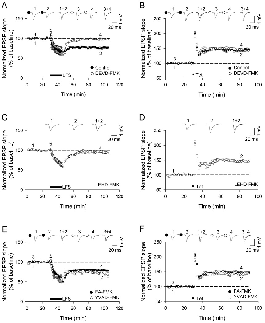
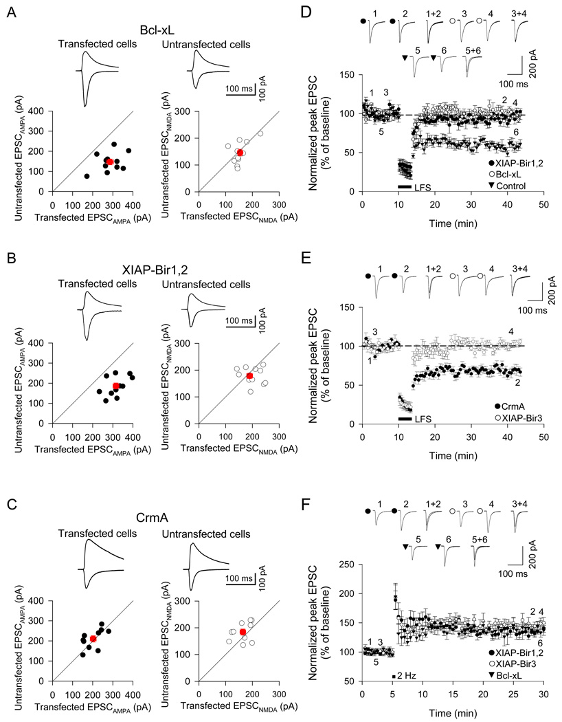
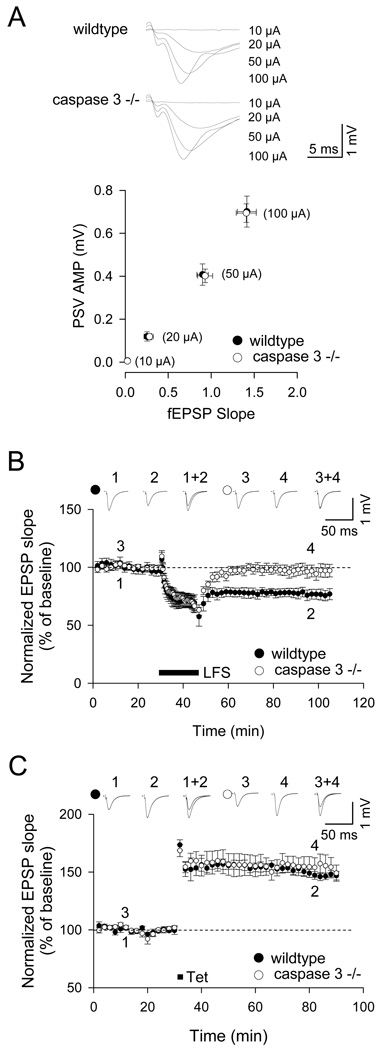
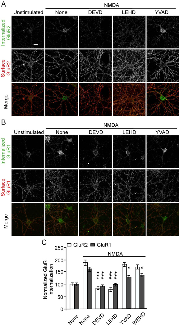
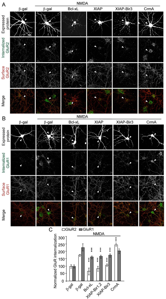
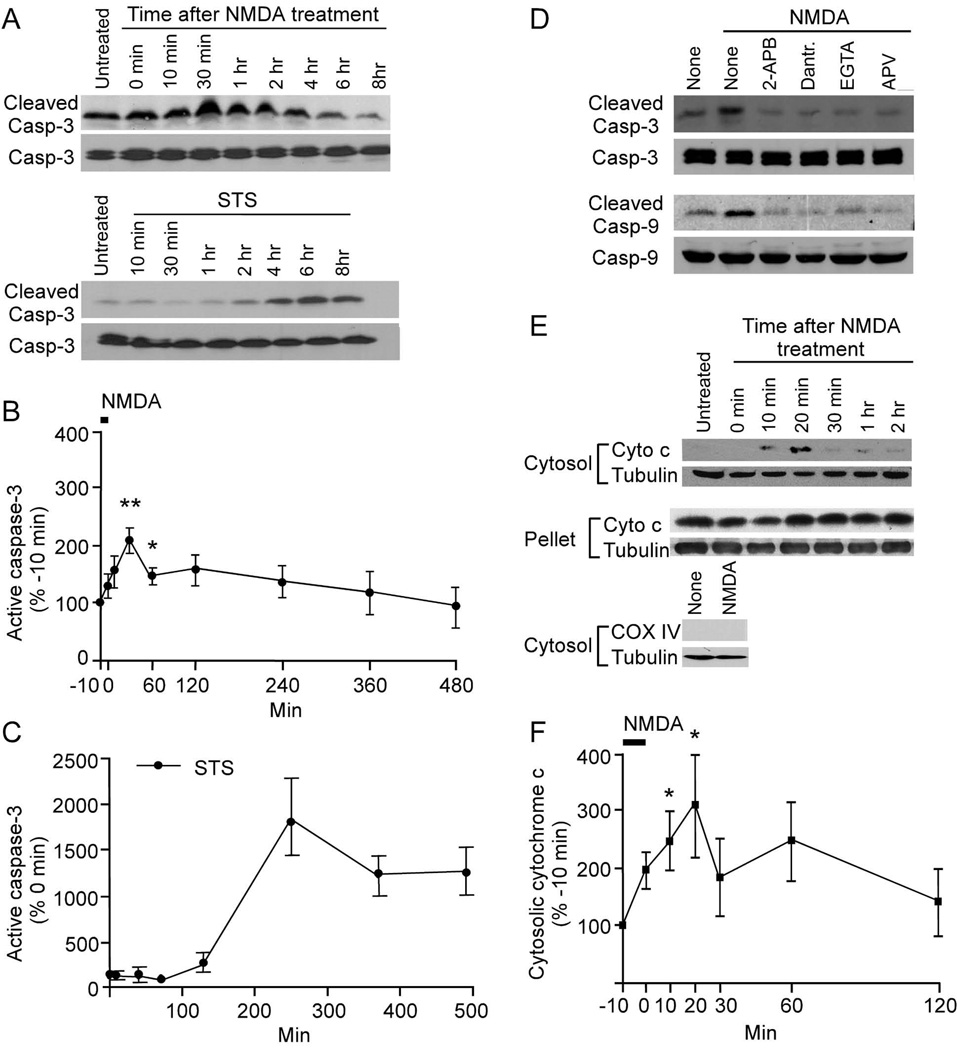
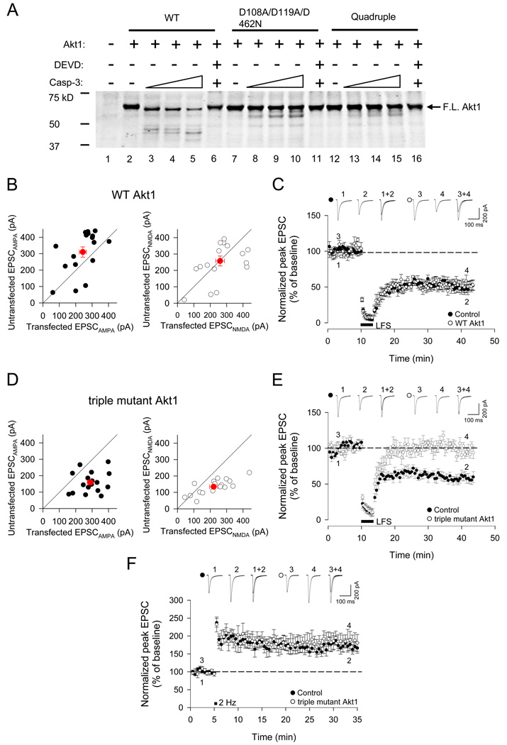
NMDA receptor-dependent synaptic modifications, such as long-term potentiation (LTP) and long-term depression (LTD), are essential for brain development and function. LTD occurs mainly by the removal of AMPA receptors from the postsynaptic membrane, but the underlying molecular mechanisms remain unclear. Here, we show that activation of caspase-3 via mitochondria is required for LTD and AMPA receptor internalization in hippocampal neurons. LTD and AMPA receptor internalization are blocked by peptide inhibitors of caspase-3 and -9. In hippocampal slices from caspase-3 knockout mice, LTD is abolished whereas LTP remains normal. LTD is also prevented by overexpression of the anti-apoptotic proteins XIAP or Bcl-xL, and by a mutant Akt1 protein that is resistant to caspase-3 proteolysis. NMDA receptor stimulation that induces LTD transiently activates caspase-3 in dendrites, without causing cell death. These data indicate an unexpected causal link between the molecular mechanisms of apoptosis and LTD.


Figures

Figure 1
Figure 1
Caspase inhibitors DEVD- and LEHD-FMK block LTD in CA1 hippocampal neurons. Acute hippocampal slices (from 3–4 week old rats) were incubated with caspase inhibitors for 2 hr before recording (DEVD-FMK, 2 µM; LEHD-FMK, 2 µM; FA-FMK, 10 µM; YVAD-FMK, 5 µM). Extracellular field EPSPs were evoked by stimulating Schaffer collateral – CA1 synapses. (A and B) Effect of DEVD-FMK on LTD and LTP, induced by low frequency stimulation (LFS) and tetanic stimulation (Tet) respectively (control, n=6; DEVD-FMK, n=6). (C and D) Effect of LEHD-FMK on LTD and LTP (n=6). (E and F) Effect of YVAD-FMK or negative control peptide FA-FMK on LTD and LTP (n=6). Pooled data and example traces are shown in all panels. Symbols and error bars indicate mean ± SEM. See also Figure S1.
Figure 2
Figure 2
Overexpression of Bcl-xL and XIAP constructs blocks LTD in CA1 neurons of organotypic hippocampal slice cultures. CA1 neurons were biolistically transfected with Bcl-xL, XIAP-Bir1,2, XIAP-Bir3 or CrmA (with GFP cotransfected as a visual marker) and recorded in whole-cell patch-clamp mode. (A–C) Pairwise analysis of the effects of Bcl-xL (11 pairs of transfected and neighboring untransfected cells), XIAP-Bir1,2 (11 pairs) or CrmA transfection (10 pairs) on basal AMPA-EPSC amplitude (left graph, recorded at a holding potential of −70mV) and NMDA-EPSC amplitude (right graph, recorded at a holding potential of +40mV) versus nearby untransfected cells (pairs of transfected and neighboring untransfected cells are individually plotted; black circles). Red symbol and error bars indicate mean ± SEM. Bcl-xL and XIAP caused an increased basal EPSCAMPA (rightward shift on graph) but had no effect on the EPSCNMDA, in comparison with neighboring untransfected cells. (D) Induction of LTD was blocked in CA1 cells overexpressing XIAP-Bir1,2 (p>0.05, n=7), and BclxL (p>0.05, n=8). LTD was intact in non-transfected CA1 neurons from organotypic hippocampal culture (p<0.01, n=6). (E) LTD induction was blocked by overexpression of XIAP-Bir3 (n=8) but not CrmA (n=8). (F) LTP was unaffected by overexpression of Bcl-xL (n=7), XIAP-Bir1,2 (n=7) or XIAP-Bir3 proteins (n=7). Graphs show mean ± SEM.
Figure 3
Figure 3
LTD is abolished in hippocampal slices from caspase-3 knockout (−/−) mice. (A) Input-output relationship in caspase-3 −/− and wildtype mice (2–4 weeks age). fEPSP slope was plotted against amplitude of presynaptic volley at four different stimulus intensities (10 µA, 20 µA, 50 µA and 100 µA). Graph indicates mean ± SEM (p>0.05, n=16). (B) Low frequency stimulation (LFS, 1 Hz, 15 min) failed to induce LTD in caspase-3 −/− slices (p<0.01, n=6). (C) Magnitude of LTP induced by tetanic stimulation (Tet) was not different between caspase-3 −/− and wildtype slices (p>0.05, n=6). See also Figure S2.
Figure 4
Figure 4
Caspase inhibitors DEVD- and LEHD-FMK block NMDA-induced AMPA receptor internalization. Antibody-feeding internalization assay for endogenous GluR2 (A) and GluR1 (B) in hippocampal neurons stimulated with NMDA (50 µM for 10 min). Neurons were pretreated with various caspase inhibitors (5 µM) as indicated. A and B show double-label immunostaining for internalized GluR (green, first row) and surface-remaining GluR (red, second row), and merge (in color, third row). Individual channels are shown in grayscale. Quantitation in C shows internalization index (integrated fluorescence intensity of internalized GluR / integrated fluorescence intensity of internalized GluR plus surface GluR) normalized to unstimulated untreated cells (Lee et al., 2002). For all quantitations, n=15 neurons for each group. *p<0.05, ***p<0.001, compared to NMDA treated cells transfected with µ-gal. The graph shows mean ± SEM. Scale bar, 20 µm. See also Figure S3.
Figure 5
Figure 5
Anti-apoptotic proteins inhibit AMPA receptor internalization. Cultured hippocampal neurons (DIV14) were transfected with Bcl-xL, XIAP-Bir1,2, XIAP-Bir3 or CrmA, or control β-gal. At 2–4 days after transfection, neurons were treated with NMDA as indicated, and GluR2 (A) and GluR1 internalization (B) was measured by the antibody-feeding internalization assay, as in Figure 4. A and B show immunostaining for the transfected protein (upper row), internalized GluR (second row), surface-remaining GluR (third row), and merge of internalized and surface GluR (bottom row). All individual channels of this triple labeling experiment are shown in grayscale. Arrowheads mark the cell body of transfected cells. Note that the cell body of a Bcl-xL transfected neuron in (A) lies adjacent to an untransfected neuron. Histograms show internalization index for GluR normalized to unstimulated control-transfected neurons (C). n=15–30 neurons for each group. ***p<0.001 (compared to NMDA stimulated β-gal control). The graph shows mean ± SEM. Scale bar, 20 µm. See also Figure S4.
Figure 6
Figure 6
Transient activation of caspase-3 in neurons by NMDA. (A, D, E) Immunoblots showing cleaved (active) caspase-3 and caspase-9, and cytosolic cytochrome c in cortical neuronal cultures (DIV18) at various times after treatment with NMDA (50 µM for 10 min) or staurosporine (STS, 1 µM throughout the experimental period). In (D), neurons were preincubated as indicated with 2-APB (30 µM), dantrolene (100 µM), DL-APV (100 µM) or EGTA (5 mM) for 15 min before NMDA treatment, and were immunoblotted 30 min after NMDA stimulation for active (cleaved) and total caspase-3,-9; and cytochrome c in the cytosolic and pellet fractions. (B, C, F) Quantitation (mean ± SEM) of immunoblot band intensities normalized to values before treatment (NMDA was applied from −10 to 0 min; STS treatment began at 0 min); n=3 for each time point. See also Figure S5.
Figure 7
Figure 7
Overexpression of caspase-3-resistant triple mutant of Akt1 (D108A/D119A/D462N) blocks LTD in CA1 neurons of organotypic hippocampal slice cultures. (A) Cleavage of wildtype or mutant Akt1 proteins by caspase-3 in vitro. In vitro translated WT Akt1, D108A/D119A/D462N triple mutant or Quadruple mutant were incubated with increasing amounts of recombinant active caspase-3 (10, 20, and 40 ng) at 37°C for 18 h. The caspase-3 inhibitor DEVD-fmk was either omitted or included in the reactions containing 40 ng caspase-3. Position of full-length Akt1 is indicated. (B) Pairwise analysis of wildtype Akt1 (Akt1) transfected cells and nearby untransfected cells on basal AMPA receptor-mediated EPSCs (left panel, EPSCAMPA, recorded at a holding potential of −70mV) and NMDA receptor-mediated EPSCs (right panel, EPSCNMDA, recorded at +40mV). Red symbols show mean ± SEM. (C) Overexpression of WT Akt1 had no effect on LTD compared to neighboring untransfected cells (p<0.01, n=6). (D) Pairwise analysis of Akt1triple mutant (D108A/D119A/D462N)-transfected cells and nearby untransfected cells on basal AMPA receptor-mediated EPSCs and NMDA receptor-mediated EPSCs. (E) Induction of LTD was blocked in Akt1 triple mutant transfected cells (p>0.05, n=6) but LTD was intact in neighboring untransfected cells. (F) LTP was unaffected by overexpression of Akt1 triple mutant. Graphs show mean ± SEM. See also Figure S6.

Comment in

References

    1. Anderson WWaC, GL The LTP Program: a data acquisition program for on-line analysis of long-term potentiation and other synaptic events. J Neurosci Methods. 2001;108:71–83. - PubMed
    1. Bachelder RE, Ribick MJ, Marchetti A, Falcioni R, Soddu S, Davis KR, Mercurio AM. p53 inhibits alpha 6 beta 4 integrin survival signaling by promoting the caspase 3-dependent cleavage of AKT/PKB. J Cell Biol. 1999;147:1063–1072. - PMC - PubMed
    1. Barry MF, Vickery RM, Bolsover SR, Bindman LJ. Intracellular studies of heterosynaptic long-term depression (LTD) in CA1 of hippocampal slices. Hippocampus. 1996;6:3–8. - PubMed
    1. Beurel E, Jope RS. The paradoxical pro- and anti-apoptotic actions of GSK3 in the intrinsic and extrinsic apoptosis signaling pathways. Progress in neurobiology. 2006;79:173–189. - PMC - PubMed
    1. Campbell DS, Holt CE. Apoptotic pathway and MAPKs differentially regulate chemotropic responses of retinal growth cones. Neuron. 2003;37:939–952. - PubMed

Publication types

MeSH terms