Review
. 2010 Apr;4(4):226-37.
doi: 10.1186/1479-7364-4-4-226.

From DNA to proteins via the ribosome: structural insights into the workings of the translation machinery

Affiliations
Review

From DNA to proteins via the ribosome: structural insights into the workings of the translation machinery

Xabier Agirrezabala et al. Hum Genomics. 2010 Apr.

Abstract

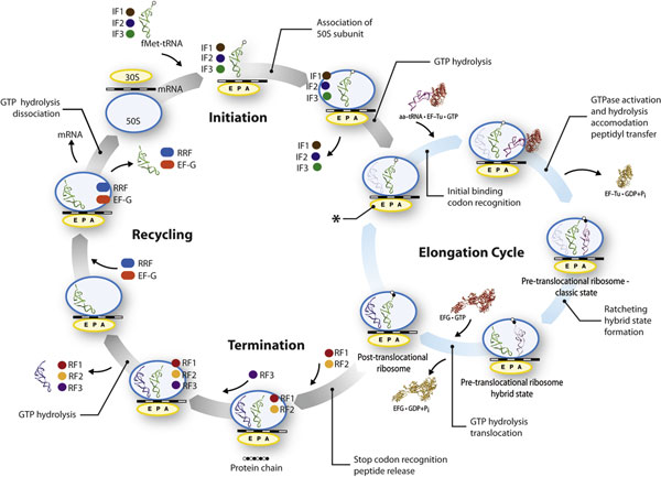
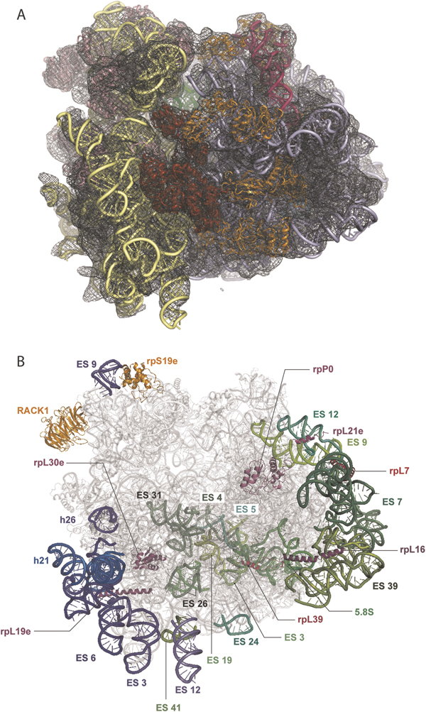
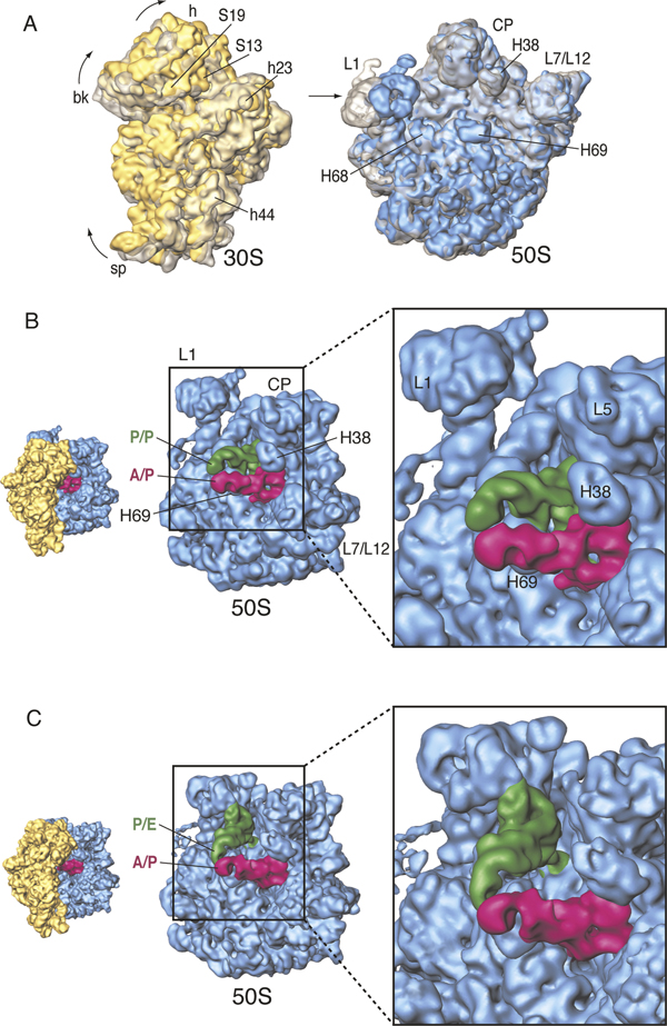
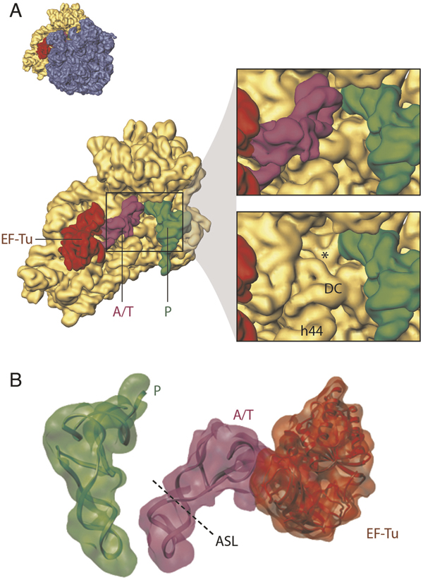
Understanding protein synthesis in bacteria and humans is important for understanding the origin of many human diseases and devising treatments for them. Over the past decade, the field of structural biology has made significant advances in the visualisation of the molecular machinery involved in protein synthesis. It is now possible to discern, at least in outline, the way that interlocking ribosomal components and factors adapt their conformations throughout this process. The determination of structures in various functional contexts, along with the application of kinetic and fluorescent resonance energy transfer approaches to the problem, has given researchers the frame of reference for what remains as the greatest challenge: the complete dynamic portrait of protein synthesis in the cell.


Figures

Figure 1
Figure 1
Schematic diagram of bacterial protein synthesis. Shown are the mains steps comprising the translation process: initiation, elongation cycle, termination and recycling. Details regarding each step are provided in the main text. For simplicity, some intermediate stages are omitted in this overview. The mRNA is depicted as a strand running horizontally along the small (30S) subunit, with alternating white and black segments, each representing one codon. The tRNAs bind at A, P and E sites. The nascent polypeptide is shown as a string of spheres. The individual structures and cartoons are not drawn to scale.
Figure 2
Figure 2
The structure of the eukaryotic ribosome derived from cryo-EM. (A) The atomic model for the yeast 80S ribosome was obtained by building on the X-ray structure of the E. coli ribosome using rRNA modelling of expansion segments, homology modelling of proteins for which there are bacterial counterparts, and placement of some extra 80S proteins whose structure is known [4]. Experimental cryo-EM densities corresponding to 40S and 60S subunits and eEF2 for Thermomyces lanuginosus, used as constraints for modelling, are shown in transparent yellow, blue and red, respectively. (B) Same as (A) but with extra proteins highlighted (orange, 40S; magenta, 60S), for which the structures are known and which could be located through cross-linking or by exhaustive computational search. (Note that helices marked rpL19e, rpL21e, rpL7, rpL16, and rpL16 have bacterial/archaeal homologues represented in the available X-ray structures for the largest portion of the proteins.) Marked are only pieces (a-helices) supported by EM density proximal to the corresponding protein and by secondary structure predictions.) Likewise, rRNA expansion segments and 5.8S rRNA are highlighted (blue, 40S; green, 60S). In (B), eEF2 has been omitted for clarity. The illustration in (A) was reproduced from Frank (2009).
Figure 3
Figure 3
Principle of cryo-EM and single-particle reconstruction. Molecules (in this case ribosomes) lying in random orientations are embedded in a thin layer of ice. Exposure to a low-dose electron beam in the transmission electron microscope produces a projection image ('electron micrograph'). A typical electron micrograph shows E. coli ribosomes as low-contrast single particles on a noisy background. After the orientations of the particles have been determined, usually by matching them with a reference, they are used to reconstruct a density map by a back-projection or a similar reconstruction algorithm. This density map is segmented into the different components (subunits, ligands), and the different components are displayed using different colours in a surface representation (bottom panel; small and large subunits are shown in yellow and blue, respectively. A- and P-site tRNAs are coloured pink and green, respectively).
Figure 4
Figure 4
Ratchet-like motion and hybrid tRNA configuration. (A) Superimposition of 30S (left) and 50S (right) subunits, as seen from the inter-subunit side. Classic-state subunits are shown in transparent grey, while the subunits of the hybrid-state ribosome are in solid yellow (30S) and blue (50S). (B) Close-up view of the 50S subunit showing the classic tRNA configuration. (C) Close-up view of the 50S subunit showing the hybrid tRNA configuration. The orientations of the subunits are shown as successive thumbnails on the left. Data reproduced from Agirrezabala et al.[37]
Figure 5
Figure 5
Decoding and aa-tRNA incorporation. (A) Close-up view of the 30S subunit showing the decoding centre (DC) region in the presence of a cognate ternary complex. In the lower panel, the tRNA is computationally removed to show the tip of helix 44, in which A1492 and A1493 flip out as the result of a cognate codon-anticodon interaction. The resulting configuration is labelled with an asterisk. The orientation of the small subunit is shown as successive thumbnails on the left. (B) Arrangement of the P-site tRNA and ternary complex. The tRNA atomic models (displayed in ribbons) were obtained by fitting of the experimental cryo-EM densities (shown as transparent) with the X-ray-derived coordinates of the tRNAs by real-space refinement.

References

    1. Acker MG, Lorsch JR. 'Mechanism of ribosomal joining during eukaryotic translation initiation'. Biochem Soc Trans. 2008;36:653–657. doi: 10.1042/BST0360653. - DOI - PubMed
    1. Dinman JD. 'The eukaryotic ribosome: Current status and challenges'. J Biol Chem. 2008;284:11761–11765. - PMC - PubMed
    1. Spahn CM, Beckmann R, Eswar N. et al. 'Structure of the 80S ribosome from Saccharomyces cerevisiae -- tRNA-ribosome and subunit-subunit interactions'. Cell. 2001;107:373–386. doi: 10.1016/S0092-8674(01)00539-6. - DOI - PubMed
    1. Taylor DJ, Devkota B, Huang AD. et al. 'Comprehensive molecular structure of the eukaryotic ribosome'. Structure. 2009;17:1591–1604. doi: 10.1016/j.str.2009.09.015. - DOI - PMC - PubMed
    1. Musters W, Boon CA, van der Sande H. et al. 'Functional analysis of transcribed spacers of yeast ribosomal DNA'. EMBO J. 1990;9:3989–3996. - PMC - PubMed

Publication types

LinkOut - more resources