Review
. 2010 Apr;4(4):250-62.
doi: 10.1186/1479-7364-4-4-250.

Hyperferritinaemia-cataract syndrome: worldwide mutations and phenotype of an increasingly diagnosed genetic disorder

Affiliations
Review

Hyperferritinaemia-cataract syndrome: worldwide mutations and phenotype of an increasingly diagnosed genetic disorder

Gunda Millonig et al. Hum Genomics. 2010 Apr.

Abstract

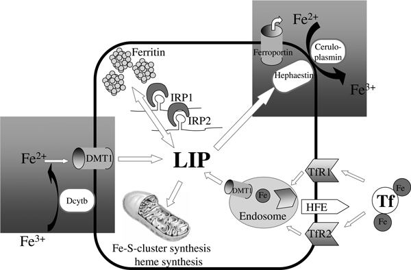
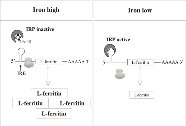
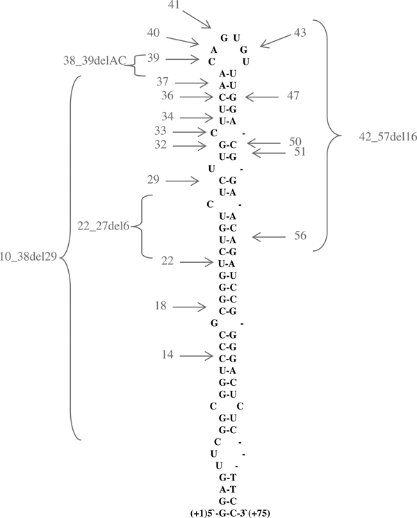
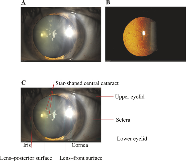
The hereditary hyperferritinaemia-cataract syndrome (HHCS) is characterised by an autosomal dominant cataract and high levels of serum ferritin without iron overload. The cataract develops due to L-ferritin deposits in the lens and its pulverulent aspect is pathognomonic. The syndrome is caused by mutations within the iron-responsive element of L-ferritin. These mutations prevent efficient binding of iron regulatory proteins 1 and 2 to the IRE in L-ferritin mRNA, resulting in an unleashed ferritin translation. This paper reviews all 31 mutations (27 single nucleotide transitions and four deletions) that have been described since 1995. Laboratory test showing hyperferritinaemia, normal serum iron and normal transferrin saturation are indicative for HHCS after exclusion of other causes of increased ferritin levels (inflammation, malignancy, alcoholic liver disease) and should prompt an ophthalmological consultation for diagnostic confirmation. Invasive diagnostics such as liver biopsy are not indicated. HHCS is an important differential diagnosis of hyperferritinaemia. Haematologists, gastroenterologists and ophthalmologists should be aware of this syndrome to spare patients from further invasive diagnosis (liver biopsy), and also from a false diagnosis of hereditary haemochromatosis followed by venesections. Patients diagnosed with HHCS should be counselled regarding the relative harmlessness of this genetic disease, with early cataract surgery as the only clinical consequence.


Figures

Figure 1
Figure 1
The role of ferritin in iron homeostasis. A virtual cell is shown, demonstrating the major cellular functions of iron uptake (TfR1 and -2, DMT1), export (ferroportin), storage (ferritin) and utilisation (eg haem synthesis). The LIP represents a chelated form of cytosolic iron that undergoes redistribution but also toxic side reactions. Hepatic hepcidin - the major systemic regulator - blocks iron export via ferroportin, while cytoplasmic IRPs coordinate cellular iron homeostasis by regulating proteins such as TfR1 and ferritin at the post-transcriptional level. Ferritin is the major iron storage protein and is mainly localised in hepatocytes and cells of the reticuloendothelial system. Connections between cellular and systemic iron metabolism are highlighted by hatched grey boxes. TfR1, transferrin receptor 1. DMT1, divalent metal transporter 1. LIP, labile iron pool. IRP, iron-regulatory protein.
Figure 2
Figure 2
Post-transcriptional control of ferritin synthesis by the IRP/IRE network. Iron depletion activates IRP-1 and -2 for binding to the IRE in the 5′-untranslated region of ferritin mRNA. This IRP-IRE interaction prevents translation of ferritin and eventually decreases ferritin protein levels. IRP, iron-regulatory protein.
Figure 3
Figure 3
Mutations causing hyperferritinaemia-cataract syndrome (HHCS) in the iron-responsive element (IRE) of L-ferritin. The IRE of L-ferritin forms a hairpin-like structure. Most of the mutations that cause HHCS discovered so far are located in the upper stem and the conserved hexanucleotide of the IRE. Single nucleotide transitions are depicted by grey arrows, and the number of the nucleotide deletions are represented by brackets. An extensive overview of all mutations can be found in Table 2.
Figure 4
Figure 4
Pathognomonic aspect of cataract in HHCS. Slit-lamp examination (a) and the retro-illumination technique (b) show the typical aspect of a star-shaped central cataract and peripheral flecks in the lens. From Millonig et al. 2009[42]. The slit-lamp is a low-power microscope combined with a high-intensity light source that can be focused to shine a narrow beam, thus allowing examination of the front parts of the eye (cornea, iris, lens). Panel (c) shows the anatomical details of (a).

References

    1. Frausto da Silva JJR, Williams RJP. The Biological Chemistry of the Elements: The Inorganic Chemistry of Life. Clarendon Press, Oxford, UK; 1991.
    1. Halliwell B, Gutteridge JMC. 'Role of free radicals and catalytic metal ions in human disease: An overview'. Methods Enzymol. 1990;186:1–85. - PubMed
    1. Hentze MW, Muckenthaler MU, Andrews NC. 'Balancing acts: Molecular control of mammalian iron metabolism'. Cell. 2004;117:285–297. doi: 10.1016/S0092-8674(04)00343-5. - DOI - PubMed
    1. Gao J, Chen J, Kramer M, Tsukamoto H. et al.'Interaction of the hereditary hemochromatosis protein HFE with transferrin receptor 2 is required for transferrin-induced hepcidin expression'. Cell Metab. 2009;9:217–227. doi: 10.1016/j.cmet.2009.01.010. - DOI - PMC - PubMed
    1. Feder JN, Gnirke A, Thomas W, Tsuchihashi Z. et al.'A novel MHC class I-like gene is mutated in patients with hereditary haemochromatosis'. Nat. Genet. 1996;13:399–408. doi: 10.1038/ng0896-399. - DOI - PubMed

Publication types