Comparative Study
. 2010 Jul;12(7):655-63.
doi: 10.1093/neuonc/nop062. Epub 2010 Feb 5.

Evaluation of retinoic acid therapy for OTX2-positive medulloblastomas

Affiliations
Comparative Study

Evaluation of retinoic acid therapy for OTX2-positive medulloblastomas

Renyuan Bai et al. Neuro Oncol. 2010 Jul.

Abstract

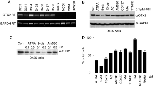
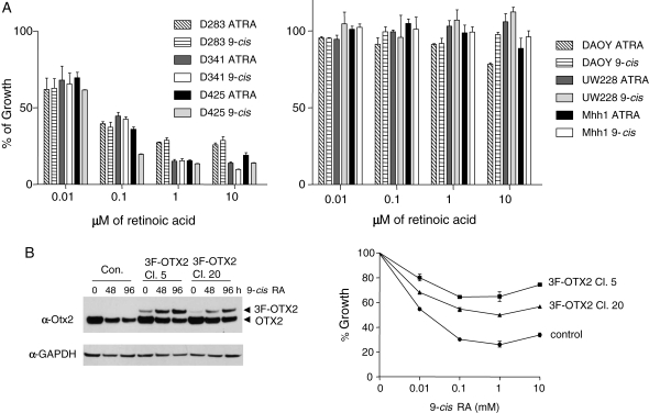
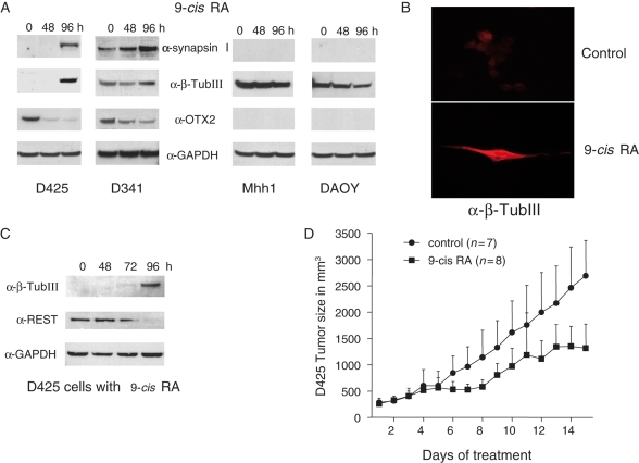
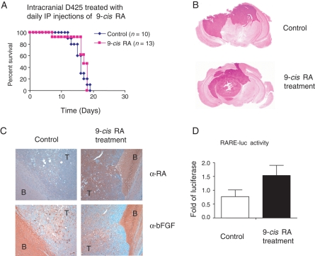
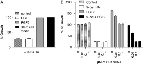
The homeobox transcription factor OTX2 plays an essential role during embryonic brain development. It is normally silenced in the adult brain, but is overexpressed by genomic amplification or other mechanisms in the majority of medulloblastomas (MBs). Retinoic acids (RAs) can suppress OTX2 expression and inhibit MB growth. In this study, 9-cis RA most potently inhibited MB cell growth. 9-cis RA functions through the downregulation of OTX2 expression, which subsequently induces neuronal differentiation of OTX2-expressing cells. Treatment with 9-cis RA reduced the growth of D425 flank xenograft tumors in mice. In an intracranial model, however, MB tumors showed resistance to 9-cis RA treatment, and we implicated fibroblast growth factor (FGF) as a potential mediator of resistance to RA therapy. These findings suggest a mechanism for RA-mediated anti-tumor effect on OTX2-positive MB cells and indicate that therapeutic targeting of OTX2 might be effective if FGF pathway-mediated resistance can be overcome.


Figures

Fig. 1.
Fig. 1.
9-cis retinoic acid (RA) suppresses OTX2 expression better than other available retinoids. (A) Expression of OTX2 in MB cell lines evaluated by RT-PCR. OTX2 or GAPDH primers were used with cDNA from the 11 MB cell lines for 28 cycles of PCR. (B) 9-cis RA suppresses OTX2 expression better than other retinoic acids. A panel of RAs at 0.1 µM was incubated with D425 cells for 48 hours. Protein level of OTX2 was analyzed by Western blotting. Equal loading was confirmed by probing with an anti-GAPDH antibody. Other retinoids tested were: ATRA, all trans-RA; 13-cis, 13-cis RA; AC, acitretin; MA, methoprene acid; TTNPB, 4-[E-2-(5,6,7,8-Tetrahydro-5,5,8,8-tetramethyl-2-naphthalenyl)-1-propenyl]benzoic acid. (C) Comparison of ATRA, 9-cis RA, and AM580 in suppressing OTX2 expression. D425 cells were incubated with 0.1 or 0.5 µM of the indicated retinoids for 48 hours. OTX2 protein level was detected by Western blotting. (D) Comparison of RAs in inhibiting the growth of D425 cells. A volume of 0.1 µM of the indicated RAs was incubated with D425 cells for 6 days with a re-addition of RAs after 3 days. Viable cells were monitored by cell proliferation assay reagent WST-1 and calculated in percentage to the DMSO control. RAHA, retinoic acid p-hydroxyanilide; GA, geranylgeranoic acid.
Fig. 2.
Fig. 2.
Suppression of MB cells by RA is found only in cells expressing OTX2, and RA sensitivity is lost with exogenous OTX2 expression. (A) Comparison of the growth inhibition from ATRA and 9-cis RA on MB cell lines. Medulloblastoma cell lines (D283, D341, D425, DAOY, UW228, and Mhh1) were incubated with DMSO (control), 0.01, 0.1, 1, or 10 µM of the indicated RA for 6 days. After 3 days, we re-added RA. Viable cells were monitored by WST-1 and calculated in percentage to the DMSO control. All the OTX2-positive cells (D283, D341, and D425) were inhibited by ATRA and 9-cis RA, while 9-cis RA generally exhibited inhibition at slightly lower concentration in D425 cells. (B) Overexpression of exogenous OTX2 reduces the growth inhibitory effect of 9-cis RA on D425 cells. OTX2 was subcloned in fusion with 3 copies of FLAG tag (3F-OTX2) and stably transfected in D425 cells. Single clones were selected by limited dilution and tested for 3F-OTX2 expression by Western blotting. D425 cells with vector alone were used as a negative control. On the anti-OTX2 Western blot, 3F-OTX2 showed a slower migration pattern compared with that of the endogenous OTX2 protein. Clone 5 (Cl. 5), Clone 20 (Cl. 20), and control cells were incubated with DMSO, 0.01, 0.1, 1, or 10 mM 9-cis RA for 96 hours. Viable cells were measured by WST-1 and graphed in percentage to the DMSO control (0 hour).
Fig. 3.
Fig. 3.
9-cis RA induces neuronal differentiation and reduces growth in a flank xenograft tumor. (A) Induction of neuronal differentiation markers in OTX2-positive MB cells by 9-cis RA. D425, D341, Mhh1, and DAOY cells were incubated with 1 µM 9-cis RA up to 96 hours. Control (0 hour) is done with incubation with DMSO for 96 hours. Western blots revealed the induction of synapsin I and β-tubulin III (β-TubIII). (B) β-TubIII immunofluorescence staining of D425 cells untreated or treated by 9-cis RA. D425 cells were incubated with 2 µM 9-cis RA for 10 days and stained with α-β-TubIII (Tuj1) antibody and Texas Red-conjugated goat anti-mouse secondary antibody. (C) 9-cis RA suppresses REST and induces β-tubulin III (β-TubIII). D425 cells were incubated with 1 µM 9-cis RA in a time course of 48, 72, and 96 hours. Western blotting reveals that 9-cis RA will suppress REST and induce β-tubulin III expression. (D) Treatment of 9-cis RA reduces flank D425 xenograft tumors. Nude mice were injected with 4 million D425 cells. After 7 days, the mice were treated with 9-cis RA dissolved in DMSO via intraperitoneal injection. Mice in the control group were treated with DMSO alone. The daily treatment of the first 7 days was paused for 1 day and resumed in the following days. The tumor size and the weight of mice were measured. The P-value at day 15 is .0006.
Fig. 4.
Fig. 4.
9-cis RA does not extend survival in mice with intracranial MB tumors. (A) No survival advantage in treatment by 9-cis RA. One million D425 cells were intracranially injected into mice and treated by IP injection of 50 µL of 9-cis RA dissolved in DMSO (n = 13) or treated with DMSO as control (n = 10). The daily treatment of the first 6 days was followed by a pause of 2 days and 5 additional daily injections. (B) Sections of H&E staining of mouse brains with or without the treatment by 9-cis RA. The darker nucleus-rich area represents the tumor formed by D425 cells. (C) α-RA and α-bFGF staining of mouse brains with or without the 9-cis RA treatment. Brain slides shown in (B) were stained with α-RA or α-bFGF and the positive staining was visualized by DAB colored in brown with hematoxylin in blue as counterstaining. Slides of serial cut of 10 µm apart were used and shown in ×10 magnification. Compared with the control, the brain with 9-cis RA treatment revealed an increased level of RA and generally unchanged bFGF expression. T, tumor; B, brain. (D) Luciferase reporter assay with the brain lysates. RARE-luc reporter construct with RA-responsive element were transfected in P19 cells and incubated with lysates of brain samples treated with or without 9-cis RA. The relative luciferase activity was graphed as the fold increase compared with the control. Three brain samples were used in each group.
Fig. 5
Fig. 5
FGF2 renders the MB cells insensitive to RA treatment. (A) FGF-2 was capable of overriding the growth-inhibitory effect of 9-cis RA on D425 cells. D425 cells were incubated with 20 ng/mL of EGF, 10 ng/mL of FGF-2 in DMEM media containing 10% FBS or stem/progenitor cell media for 96 hours in the presence of 1 µM 9-cis RA. Viable cells were determined by cell proliferation assay reagent WST-1 and graphed in percentage to the control that was cultured without 9-cis RA. (B) FGFR inhibitor PD173074 restored the RA-sensitivity of D425 cells. D425 cells were incubated for 96 hours with the indicated combinations of 9-cis RA (1 µM) and FGF2 (10 ng/mL) in the presence of the FGFR inhibitor PD173074 at the concentration from 0 to 1 µM. Viable cells were determined by WST-1 and graphed in percentage to the control that was cultured without 9-cis RA, FGF2, and PD173074.

References

    1. Kleihues P, Louis DN, Scheithauer BW, et al. The WHO classification of tumors of the nervous system. J Neuropathol Exp Neurol. 2002;61:215–225. - PubMed
    1. Palmer SL, Reddick WE, Gajjar A. Understanding the cognitive impact on children who are treated for medulloblastoma. J Pediatr Psychol. 2007;32:1040–1049. - PubMed
    1. Yokota N, Mainprize TG, Taylor MD, et al. Identification of differentially expressed and developmentally regulated genes in medulloblastoma using suppression subtraction hybridization. Oncogene. 2004;23:3444–3453. - PubMed
    1. Boon K, Eberhart CG, Riggins GJ. Genomic amplification of orthodenticle homologue 2 in medulloblastomas. Cancer Res. 2005;65:703–707. - PubMed
    1. Di C, Liao S, Adamson DC, et al. Identification of OTX2 as a medulloblastoma oncogene whose product can be targeted by all-trans-retinoic acid. Cancer Res. 2005;65:919–924. - PubMed

Publication types

MeSH terms