. 2011 Feb;10(2):M110.001131.
doi: 10.1074/mcp.M110.001131. Epub 2010 May 28.

Proteomics profiling of Madin-Darby canine kidney plasma membranes reveals Wnt-5a involvement during oncogenic H-Ras/TGF-beta-mediated epithelial-mesenchymal transition

Affiliations

Proteomics profiling of Madin-Darby canine kidney plasma membranes reveals Wnt-5a involvement during oncogenic H-Ras/TGF-beta-mediated epithelial-mesenchymal transition

Yuan-Shou Chen et al. Mol Cell Proteomics. 2011 Feb.

Abstract

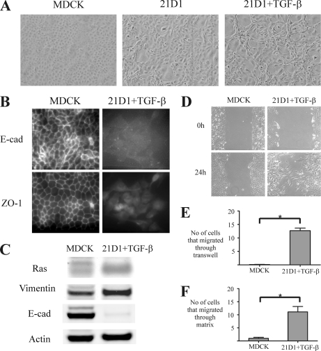
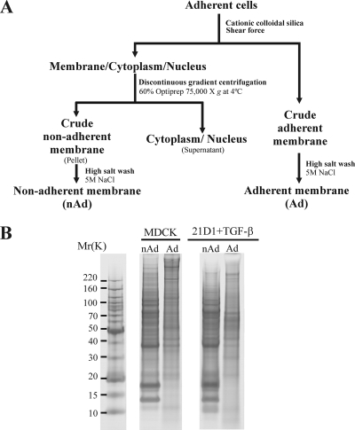
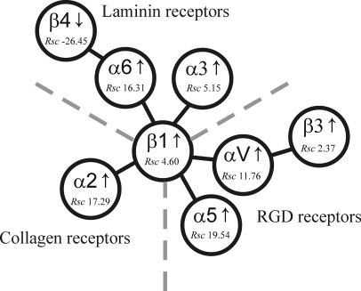
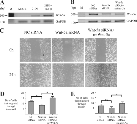
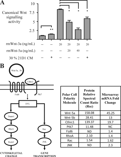
Epithelial-mesenchymal transition (EMT) describes a process whereby polarized epithelial cells with restricted migration transform into elongated spindle-shaped mesenchymal cells with enhanced motility and invasiveness. Although there are some molecular markers for this process, including the down-regulation of E-cadherin, our understanding of plasma membrane (PM) and associated proteins involved in EMT is limited. To specifically explore molecular alterations occurring at the PM, we used the cationic colloidal silica isolation technique to purify PM fractions from epithelial Madin-Darby canine kidney cells during Ras/TGF-β-mediated EMT. Proteins in the isolated membrane fractions were separated by one-dimensional SDS-PAGE and subjected to nano-LC-MS/MS-based protein identification. In this study, the first membrane protein analysis of an EMT model, we identified 805 proteins and determined their differential expression using label-free spectral counting. These data reveal that Madin-Darby canine kidney cells switch from cadherin-mediated to integrin-mediated adhesion following Ras/TGF-β-mediated EMT. Thus, during the EMT process, E-cadherin, claudin 4, desmoplakin, desmoglein-2, and junctional adhesion molecule A were down-regulated, whereas integrins α6β1, α3β1, α2β1, α5β1, αVβ1, and αVβ3 along with their extracellular ligands collagens I and V and fibronectin had increased expression levels. Conspicuously, Wnt-5a expression was elevated in cells undergoing EMT, and transient Wnt-5a siRNA silencing attenuated both cell migration and invasion in these cells. Furthermore, Wnt-5a expression suppressed canonical Wnt signaling induced by Wnt-3a. Wnt-5a may act through the planar cell polarity pathway of the non-canonical Wnt signaling pathway as several of the components and modulators (Wnt-5a, -5b, frizzled 6, collagen triple helix repeat-containing protein 1, tyrosine-protein kinase 7, RhoA, Rac, and JNK) were found to be up-regulated during Ras/TGF-β-mediated EMT.


Figures

Fig. 1.
Fig. 1.
Oncogenic H-Ras and TGF-β stimulation induces EMT in MDCK cells. A, MDCK cells transformed with oncogenic H-Ras undergo morphological alterations from round cobblestone-like cells with tight cell-cell adhesion to long spindle-shaped cells with reduced cell contact with neighboring cells. Stimulation with TGF-β further elongates cell shape and increases scattering. B, immunofluorescence microscopy reveals strong positive staining of epithelial cell junction proteins E-cadherin (E-cad) and ZO-1 between borders of MDCK cells. In contrast, 21D1 cells stimulated with TGF-β exhibit loss of E-cadherin and ZO-1 localization and protein expression. C, MDCK cells undergoing oncogenic Ras- and TGF-β-induced EMT show diminished expression of the epithelial marker E-cadherin and increased expression of the mesenchymal marker vimentin by Western immunoblotting. D, the wound healing assay reveals that 21D1 cells have increased cell migration compared with MDCK cells as individual cells leave the front to migrate into the wounded area. E, 21D1 cells exhibit elevated migration in the Transwell migration assay as a significant (*) number of 21D1 cells move through the membrane filter toward the lower chamber (n = 3). F, a significant number of 21D1 cells relative to MDCK cells penetrated through the collagen-Matrigel matrix, demonstrating their enhanced cell invasion during EMT (n = 2). Error bars represent mean ± S.D. Significance is determined by a p value ≤0.05 using the Student's t test.
Fig. 2.
Fig. 2.
Plasma membrane purification. A, experimental work flow outlining purification of Ad and nAd membranes from MDCK and 21D1 cells. Membrane protein isolation and purification are based on the cationic colloidal silica method (26) and a modified version (31). B, SDS-PAGE protein gel banding patterns of Ad and nAd isolated membrane fractions. Gel lanes were excised and trypsinized, and extracted peptides were subjected to LC-MS/MS protein identification.
Fig. 3.
Fig. 3.
Integrin regulation during Ras/TGF-β-mediated EMT. Integrin α6β4, which binds laminin 5, was down-regulated during EMT, whereas other laminin-binding integrins, including α6β1 and α3β1, had elevated expression levels. The major collagen-binding integrin, α2β1, was up-regulated during the EMT process. Increased expression levels of integrin RGD receptors that bind fibronectin and vitronectin, namely integrins α5β1, αVβ1, and αVβ3, were detected during plasma membrane proteomics profiling. Rsc values indicate relative abundance based on label-free spectral counting (see “Experimental Procedures”). Figure was adapted from Hynes (52).
Fig. 4.
Fig. 4.
siRNA silencing of Wnt-5a attenuates 21D1 cell migration and invasion. A, transcript expression of Wnt-5a was not detected in MDCK cells by semiquantitative RT-PCR; however, transformation with H-Ras and stimulation with TGF-β induces Wnt-5a expression. B, expression of Wnt-5a in 21D1 cells is attenuated by RNA interference using siRNA duplexes targeting Wnt-5a. The addition of scrambled negative control (NC) siRNA did not affect Wnt-5a expression. Expression of Wnt-5a was reduced in 21D1 cells using transient siRNA transfection, and cell migration was assessed using the wound healing (C) and Transwell migration (D) assays. Both assays show that cell migration is significantly impeded when expression of Wnt-5a is reduced. However, cell migration is restored and even elevated with the addition of rm Wnt-5a (n = 2). E, the ability of 21D1 cells to invade through the matrix is decreased when expression of Wnt-5a is reduced using siRNA duplexes, although this reduction is not statistically significant (**). The invasive nature of 21D1 cells is rescued with the addition of recombinant Wnt-5a (n = 2). Error bars represent mean ± S.D. Significance is determined by a p value ≤0.05 using the Student's t test.
Fig. 5.
Fig. 5.
Wnt-5a expression during EMT represses canonical Wnt signaling. A, canonical Wnt signaling activity was examined using the T-cell factor-luciferase reporter assay under different culture conditions. 21D1 cells cultured in 30% conditioned medium (CM) showed no significant change (**) in canonical Wnt signaling activity. Addition of rm Wnt-3a stimulates canonical Wnt signaling; however, this activity is significantly (*) repressed following the addition of 20 ng/ml and to a greater extent by 40 ng/ml recombinant Wnt-5a. In a similar fashion, addition of 30% conditioned medium from 21D1 cells significantly attenuates canonical Wnt signaling (n = 2). Error bars represent mean ± S.D. Significance is determined by a p value ≤0.05 using the Student's t test. B, the non-canonical Wnt signaling PCP pathway may be activated during Ras/TGF-β-induced EMT. Several upstream components, core components, downstream effectors, and PCP modulators were revealed to be up-regulated in our integrated proteomics and transcriptomics EMT studies. Cthrc1, collagen triple helix repeat-containing protein 1; Fzd6, frizzled 6; Ptk7, tyrosine-protein kinase 7; DVL, dishevelled; ND, not detected; NC, no change. Pathway interactions are based on information obtained from the Kyoto Encyclopedia of Genes and Genomes database (http://www.genome.jp/kegg/pathway.html).

References

    1. Kang Y., Massagué J. (2004) Epithelial-mesenchymal transitions: twist in development and metastasis. Cell 118, 277–279 - PubMed
    1. Thiery J. P. (2002) Epithelial-mesenchymal transitions in tumour progression. Nat. Rev. Cancer 2, 442–454 - PubMed
    1. Thiery J. P. (2003) Epithelial-mesenchymal transitions in development and pathologies. Curr. Opin. Cell Biol. 15, 740–746 - PubMed
    1. Hugo H., Ackland M. L., Blick T., Lawrence M. G., Clements J. A., Williams E. D., Thompson E. W. (2007) Epithelial-mesenchymal and mesenchymal-epithelial transitions in carcinoma progression. J. Cell. Physiol. 213, 374–383 - PubMed
    1. Bailey J. M., Singh P. K., Hollingsworth M. A. (2007) Cancer metastasis facilitated by developmental pathways: sonic hedgehog, Notch, and bone morphogenic proteins. J. Cell. Biochem. 102, 829–839 - PubMed

Publication types