. 2010 Sep 16;116(11):1857-66.
doi: 10.1182/blood-2009-12-256461. Epub 2010 May 28.

The galactocerebrosidase enzyme contributes to the maintenance of a functional hematopoietic stem cell niche

Affiliations

The galactocerebrosidase enzyme contributes to the maintenance of a functional hematopoietic stem cell niche

Ilaria Visigalli et al. Blood. .

Abstract

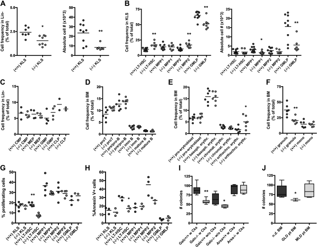
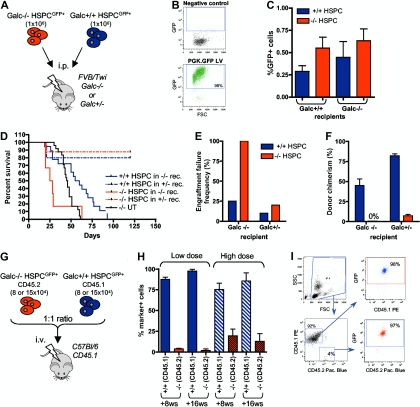
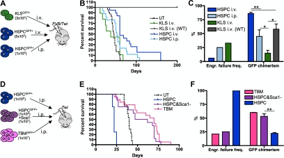
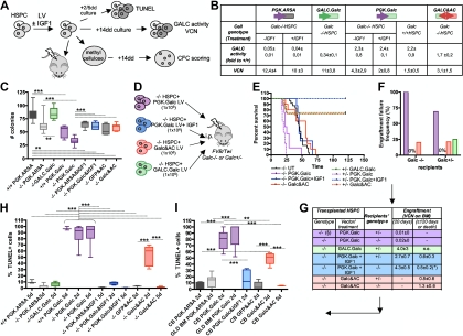
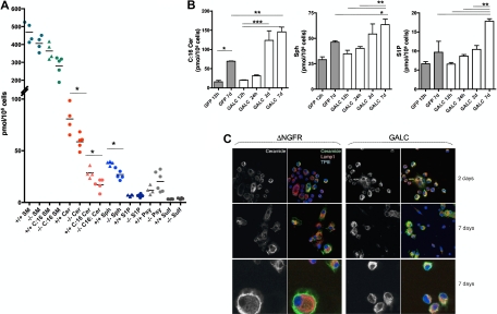
The balance between survival and death in many cell types is regulated by small changes in the intracellular content of bioactive sphingolipids. Enzymes that either produce or degrade these sphingolipids control this equilibrium. The findings here described indicate that the lysosomal galactocerebrosidase (GALC) enzyme, defective in globoid cell leukodystrophy, is involved in the maintenance of a functional hematopoietic stem/progenitor cell (HSPC) niche by contributing to the control of the intracellular content of key sphingolipids. Indeed, we show that both insufficient and supraphysiologic GALC activity-by inherited genetic deficiency or forced gene expression in patients' cells and in the disease model-induce alterations of the intracellular content of the bioactive GALC downstream products ceramide and sphingosine, and thus affect HSPC survival and function and the functionality of the stem cell niche. Therefore, GALC and, possibly, other enzymes for the maintenance of niche functionality and health tightly control the concentration of these sphingolipids within HSPCs.


Figures

Figure 1
Figure 1
Phenotype and clonogenic potential of Galc−/− HSPCs. (A-F) Immunophenotypic characterization of the BM of Galc−/− mice. Frequency and absolute cell number (#) of c-kit+ Sca1+ cells within lineage (Lin) BM cells (KLS; A), and of long-term HSC (LT-HSC), MPP1, MPP2, and GMLP (within the KLS fraction) of −/− and +/+ mice (B). Frequency of lymphoid and myeloid progenitors within Lin cells (C) of wild-type and mutant mice. Lymphoid (D), erythroid (E), and myeloid (F) differentiation in −/− and +/+ BM. Each dot represents a pool of 3 mice in panels A through C and a single mouse in panels D through F; means are shown. For details on the gating strategy, please see supplemental Table 1. (G-H) Frequency of proliferating cells at Hoechst staining (G) and of apoptotic annexin V positive cells (H) within KLS, LT-HSC, MPP1, MPP2, and GMLP from +/+ and −/− mice. Each dot represents a pool of 3 mice; means are shown. (I-J) Number (#) of colonies retrieved from colony-forming cell assays performed with murine (I) and human (J) HSPCs from wild-type (Galc+/+ and Arsa+/+), Galc−/− and Arsa −/− mice, and from normal donors' (nd) and GLD/MLD patients' BM. Before plating, murine HSPCs were (w) or were not (w/o) prestimulated with a standard cytokine cocktail for 12 hours. n ≥ 10 (murine) and n ≥ 3 (human). *P < .05, **P < .01 on 1-way ANOVA.
Figure 2
Figure 2
In vivo functional characterization of Galc−/− HSPCs. (A) Experimental scheme for panels D through F. (B) Representative cytofluorimetric dot plots of GFP-transduced HSPCs and their negative control. (C) Frequency of GFP positive cells in the BM of FVB/Twi Galc−/− and +/+ recipients 20 hours after the transplantation of GFP+ Galc−/− and +/+ HSPCs (n = 4 for each group). (D) Survival curve of Galc−/− and +/− recipient mice (rec.) after the intraperitoneal transplantation of Galc +/+ or −/− HSPCs. (E-F) Engraftment failure frequency (E; defined as percent of animals showing a survival less than or equal to 21 days postirradiation, and/or less than or equal to 1% donor cells in the BM at death or at 120 postnatal days) and donor chimerism (% of GFP+ cell in the BM) at 120 days after transplantation or at mice death (F), measured in the same experimental groups as in panel D. Average values ± SD are reported. (G) Experimental scheme for competitive transplantation experiments (H-I). (H) Engraftment of Galc+/+ CD45.1+ and of Galc−/− CD45.2+ cells in peripheral blood of wild-type CD45.1 mice transplanted with HSPCs measured at 8 and 16 weeks (ws) after the transplantation (high or the low dose, see text for details; n = 6 for each group). Average values ± SD are reported. (I) Representative plots from cytofluorimetric analysis showing engraftment of CD45.1 and CD45.2 cells on peripheral blood of mice receiving competitive transplantation and GFP expression in each of the 2 populations.
Figure 3
Figure 3
Functional characterization of the Galc−/− niche. (A,D) Experimental schemes. (B,E) Survival curves of Galc−/− mice (FVB/Twi in panel B and Twi in panel E) after the intravenous or intraperitoneal transplantation of GFP+ Galc+/+ HSPC or KLS or total BM (TBM) or HSPC plus GFP Sca1 BM cells, as indicated. KLS intravenous (WT): KLS transplanted intravenously in wild-type recipients. n ≥ 3 in each group. (C,F) Engraftment failure frequency and donor chimerism measured in the same experimental groups as in panels B and E. Average values ± SD are reported. *P < .05, **P < .01 on 1-way ANOVA.
Figure 4
Figure 4
Characterization of the diverse phenotypes associated to alterations of GALC expression in murine and human HSPCs. (A,D) Experimental schemes. (B) GALC activity (measured as fold to mock-transduced Galc+/+ cells) and LV copy number (VCN) measured on the in vitro progeny of murine Galc−/− and +/+ HSPCs transduced with the indicated LV and exposed or not to IGF1 after transduction. n ≥ 3 in each group. (C) Number (#) of colonies retrieved from colony-forming cell assays performed with murine HSPCs obtained from Galc−/− and +/+ mice after transduction with the specified LV and exposed to IGF1 after transduction, if indicated. n ≥ 3 in each group. (E) Survival curves of FVB/Twi Galc−/− and +/− mice after the intraperitoneal transplantation of Galc−/− HSPCs transduced with the indicated vectors and exposed to IGF1 after transduction, if indicated. n ≥ 3 in each group. (F-G) Engraftment failure frequency (F) and transduced cell engraftment (VCN in the BM; G) at 20 and at 120 days after transplantation or at death as in (*), measured in the same experimental groups as in panel E. (§) Similar results were obtained with +/+HSPCs. (H-I) TUNEL assay performed on murine (H) and human (I) HSPCs obtained from Galc−/− and +/+ mice and from normal donors (nd) CB and GLD patients' BM, 2 and 5 days (d) after transduction with the specified LV and exposed to IGF1 after transduction, if indicated. Percentage of TUNEL+ nuclei over the total number of nucleated cells is reported (≥ 8 fields and ≥ 100 cells were counted per condition). n ≥ 5 individual experiments per condition, average values ± SD are reported. *P < .05, **P < .01, ***P < .001 on 1-way ANOVA.
Figure 5
Figure 5
Sphingolipid profile of murine HSPCs. (A) Total sphingolipid content (pmol/106 cells) of Galc−/− and +/+ murine HSPCs. Each dot represents an individual sample; means are shown. (B) Intracellular content of C:16 Cer (left panel), Sph (middle panel), and S1P (right panel), in GALC or GFP LV transduced −/− HSPCs at 12 to 24 hours (h), 2 and 7 days (d) after gene transfer (n ≥ 3). *P < .05, **P < .01, ***P < .001 on 1-way ANOVA. (C) Representative images obtained after immunofluorescence staining for ceramide and for the lysosomal marker Lamp1 performed on human monocytes (U937 cells) transduced with GALC and control DeltaNGFR (DNGFR) LV 2 and 7 days after transduction. Magnification 100× in the 2 top rows and 200× in the bottom row.

References

    1. Suzuki K. Twenty five years of the “psychosine hypothesis”: a personal perspective of its history and present status. Neurochem Res. 1998;23(3):251–259. - PubMed
    1. Galbiati F, Basso V, Cantuti L, et al. Autonomic denervation of lymphoid organs leads to epigenetic immune atrophy in a mouse model of Krabbe disease. J Neurosci. 2007;27(50):13730–13738. - PMC - PubMed
    1. Katayama Y, Battista M, Kao WM, et al. Signals from the sympathetic nervous system regulate hematopoietic stem cell egress from bone marrow. Cell. 2006;124(2):407–421. - PubMed
    1. Hannun YA, Obeid LM. Principles of bioactive lipid signalling: lessons from sphingolipids. Nat Rev Mol Cell Biol. 2008;9(2):139–150. - PubMed
    1. Chen YQ, Wenger DA. Galactocerebrosidase from human urine: purification and partial characterization. Biochim Biophys Acta. 1993;1170(1):53–61. - PubMed

Publication types

MeSH terms