. 2010 Oct;38(19):6418-32.
doi: 10.1093/nar/gkq455. Epub 2010 May 28.

Three prime exonuclease I (TREX1) is Fos/AP-1 regulated by genotoxic stress and protects against ultraviolet light and benzo(a)pyrene-induced DNA damage

Affiliations

Three prime exonuclease I (TREX1) is Fos/AP-1 regulated by genotoxic stress and protects against ultraviolet light and benzo(a)pyrene-induced DNA damage

Markus Christmann et al. Nucleic Acids Res. 2010 Oct.

Abstract

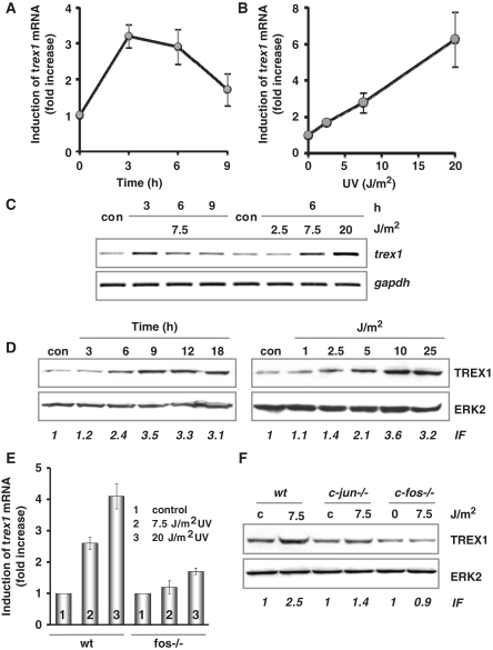
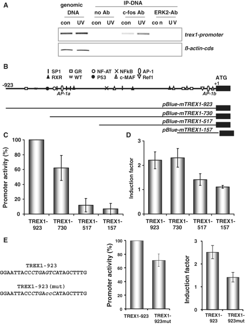
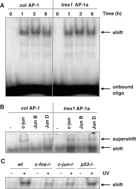
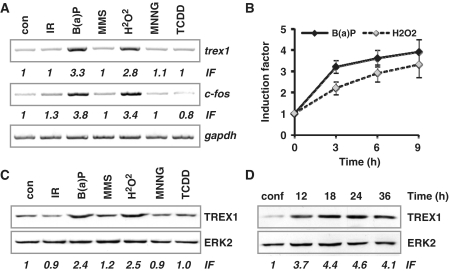
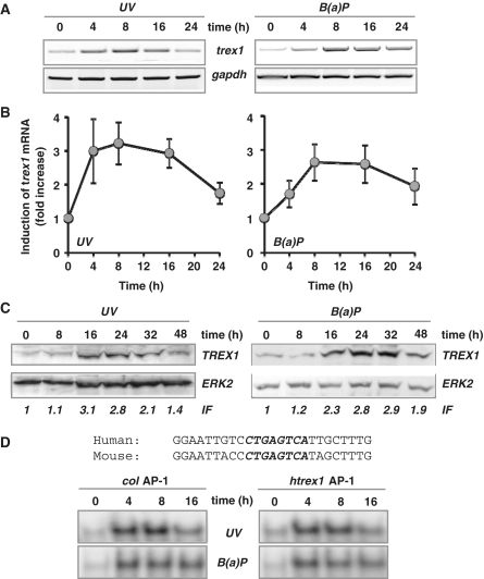
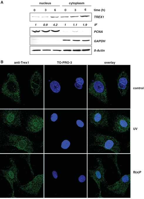
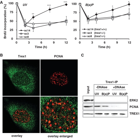
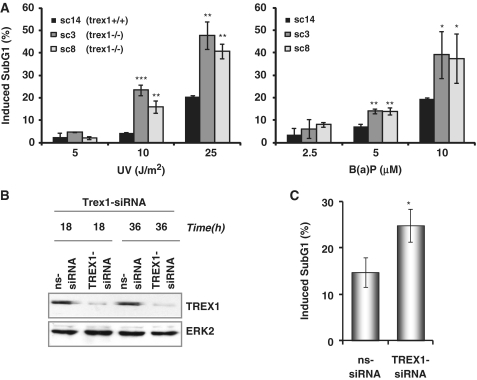
Cells respond to genotoxic stress with the induction of DNA damage defence functions. Aimed at identifying novel players in this response, we analysed the genotoxic stress-induced expression of DNA repair genes in mouse fibroblasts proficient and deficient for c-Fos or c-Jun. The experiments revealed a clear up-regulation of the three prime exonuclease I (trex1) mRNA following ultraviolet (UV) light treatment. This occurred in the wild-type but not c-fos and c-jun null cells, indicating the involvement of AP-1 in trex1 induction. Trex1 up-regulation was also observed in human cells and was found on promoter, RNA and protein level. Apart from UV light, TREX1 is induced by other DNA damaging agents such as benzo(a)pyrene and hydrogen peroxide. The mouse and human trex1 promoter harbours an AP-1 binding site that is recognized by c-Fos and c-Jun, and its mutational inactivation abrogated trex1 induction. Upon genotoxic stress, TREX1 is not only up-regulated but also translocated into the nucleus. Cells deficient in TREX1 show reduced recovery from the UV and benzo(a)pyrene-induced replication inhibition and increased sensitivity towards the genotoxins compared to the isogenic control. The data revealed trex1 as a novel DNA damage-inducible repair gene that plays a protective role in the genotoxic stress response.


Figures

Figure 1.
Figure 1.
UV light-triggered induction of TREX1. (A and B) Exponentially growing wt MEFs were exposed to 7.5 J/m2 UV for 3, 6 and 9 h (A) or exposed to 2.5, 7.5 and 20 J/m2 UV for 6 h (B). Total RNA was isolated and real-time RT-PCR was performed using trex1- and gapdh-specific primers. For quantification, the expression was normalized to gapdh and the untreated control was set to 1. Data are the mean of three independent experiments +/− SD. (C) In a different set of experiments, wt MEFs were also exposed to 7.5 J/m2 UV for 3, 6 and 9 h (left panel) or exposed to 2.5, 7.5 and 20 J/m2 UV for 6 h (right panel), total RNA was isolated and semi-quantitative RT-PCR was performed using trex1 or, as loading control, gapdh-specific primers (con, non-exposed control). (D) Exponentially growing wt MEFs were exposed to 7.5 J/m2 UV for different time points or exposed to different doses of UV for 9 h. Total protein extract was isolated. Immunodetection was performed using TREX1 or, as loading control, ERK2-specific antibody. Induction factor (IF) is derived from densitometric measurement of TREX1 signal and normalized to ERK2 expression (E) Exponentially growing wt and fos−/− MEFs were exposed to 7.5 or 20 J/m2 UV for 6 h. Total RNA was isolated and real-time RT-PCR was performed using trex1- and gapdh- specific primers. For quantification, the expression was normalized to gapdh and the untreated control was set to 1. Data are the mean of three independent experiments +/− SD. (F) Exponentially growing wt, c-jun−/− and c-fos−/− MEFs were exposed to 20 J/m2 UV for 9 h. Total protein extract was isolated. Immunodetection was performed using TREX1 or, as loading control, ERK2-specific antibody.
Figure 2.
Figure 2.
Characterization of the murine trex1 promoter. (A) ChIP analysis: exponentially growing wt cells were not exposed (con, control) or exposed to 20 J/m2 UV. Cells were harvested 6 h later and dealt with as described in ‘Material and Methods’ section. IP was performed using a c-Fos-specific antibody and (as negative control) ERK2-specific antibody. PCR was done using specific primers for the trex1 promoter and, as negative control, for the β-actin 5′-UTR. (B) Graphical visualization of several putative transcription factor binding sites identified using the program Patch 1.0 (www.gene-regulation.com) and display of generated promoter fragments. (C and D) A 923 bp genomic fragment 5′ of the ATG codon of trex1 and different trex1 promoter fragments were cloned into the pBlue-Topo vector and transiently transfected in exponentially growing Swiss albino 3T3 cells. (C) Basal promoter activity was determined by β-Gal assay and compared between untreated cells. The activity of the −923 fragment was set to 100. (D) UV-induced promoter activity was determined after exposure to 7.5 J/m2. Therefore, the promoter activity in UV-exposed cells was set in relation to the promoter activity in non-exposed cells resulting in fold induction. Data are the mean of three independent experiments +/− SD. (E) The putative AP-1 binding site (AP-1a as indicated under B) within the 923 bp genomic fragment containing pBlue-Topo vector was mutated via site-directed mutagenesis (left panel for sequences) and transiently transfected in exponentially growing Swiss albino 3T3 cells. Basal promoter activity and UV-induced promoter activity was determined after exposure to 7.5 J/m2. Data are the mean of three independent experiments +/− SD.
Figure 3.
Figure 3.
Identification of AP-1 binding sites within the trex1 promoter. (A) Binding of AP-1 to promoter fragments as determined by EMSA. Oligonucleotides containing either the AP-1 binding site of the collagenase promoter (col AP-1) or the AP-1 binding sites of the trex1 promoter (trex1 AP-1a) were incubated with nuclear extracts from wt cells exposed to 20 J/m2 UV light for 1, 3 or 6 h and subjected to EMSA. (B) EMSA supershift assay. Composition of the AP-1 factor bound to the col AP-1 and trex1 AP-1a oligonucleotide was analysed by the addition of specific antibodies to the reaction. (C) Oligonucleotides containing the AP-1 binding sites of the trex1 promoter (trex1 AP-1a) were incubated with nuclear extracts from wt cells, c-fos−/−, c-jun−/− and p53−/− cells, exposed to 20 J/m2 UV light for 6 h and subjected to EMSA.
Figure 4.
Figure 4.
Induction of trex1 by genotoxins. (A) Exponentially growing wt MEFs were exposed to different genotoxins: 2.5 µM B(a)P, 1 mM H2O2, 2 Gy IR, 25 µM MNNG, 0.5 mM MMS, 2 nM TCDD for 6 h. RNA was isolated and semi-quantitative RT-PCR was performed using trex1, c-fos or, as positive control, gapdh- specific primers (con, non-exposed control). IF, induction factor. (B) Exponentially growing wt MEFs were exposed to 2.5 µM B(a)P or 0.5 mM H2O2 for 3, 6 or 9 h. Total RNA was isolated and real-time RT-PCR was performed using trex1-specific primers. For quantification, the expression was normalized with gapdh and the untreated control was set to 1. Data are the mean of three independent experiments +/− SD. (C) Exponentially growing wt MEFs were exposed to different genotoxins: 2.5 µM B(a)P, 1 mM H2O2, 2 Gy IR, 25 µM MNNG, 0.5 mM MMS, 2 nM TCDD for 9 h. Total protein extract was isolated. Immunodetection was performed using TREX1 or, as loading control, ERK2-specific antibodies. IF, induction factor. (D) Cells arrested in G1 by confluency (conf) and cells harvested at different time points after reseeding were harvested, whole cell extracts were isolated and the expression of TREX1 was measured by immunodetection using TREX1 or, as loading control, ERK2-specific antibodies.
Figure 5.
Figure 5.
Induction of trex1 in human cells. (A) Exponentially growing human fibroblasts (cell line GM637) were exposed to 10 J/m2 UV or 2.5 µM B(a)P for different time points. Total RNA was isolated and semi-quantitative RT-PCR was performed using primers specific for trex1 or, as positive control, gapdh- specific primers (con, non-exposed control). (B) Exponentially growing GM637 cells were exposed to 10 J/m2 UV or 2.5 µM B(a)P for different time points. Total RNA was isolated and real-time RT-PCR was performed using primers specific for trex1 or, as positive control, gapdh-specific primers (con, non-exposed control). For quantification, the expression was normalized to gapdh and the untreated control was set to 1. Data are the mean of three independent experiments +/− SD. (C) Exponentially growing GM637 cells were exposed to 10 J/m2 UV or 2.5 µM B(a)P for different time points. Total protein extract was isolated. Immunodetection was performed using TREX1 or, as loading control, ERK2-specific antibodies. IF, induction factor. (D) Binding of AP-1 to promoter fragments as determined by EMSA. Oligonucleotides containing either the AP-1 binding site of the collagenase promoter (col AP-1) or the human trex1 promoter (htrex1 AP-1) were incubated with nuclear extracts from GM637 fibroblasts exposed to 10 J/m2 UV or 2.5 µM B(a)P for 4, 8 or 16 h and subjected to EMSA.
Figure 6.
Figure 6.
Nuclear translocation of TREX1. (A) Exponentially growing wt MEFs were non-exposed or exposed to 20 J/m2 UV for 3 and 6 h. Nuclear and cytoplasmic extracts were isolated. Immunodetection was performed using TREX1, PCNA, GAPDH or, as loading control, β-Actin-specific antibody. Induction factor (IF) is derived from densitometric measurement of TREX1 signal and normalized to β-Actin expression. (B) Exponentially growing wt MEFs were not exposed (control) or exposed to 20 J/m2 UV light or 2.5 µM B(a)P for 6 h and thereafter fixed as described. TREX1 localization was visualized by the use of a specific antibody and detected by confocal laser scanning microscopy.
Figure 7.
Figure 7.
Impact of TREX1 on DNA replication. (A) Exponentially growing TREX1 wt cells (sc14+/+) and TREX1-deficient cells (sc3−/− and sc8−/−) were exposed to 7.5 J/m2 UV (left panel) or 2.5 µM B(a)P (right panel). Different time points later DNA replication was measured by incorporation of BrdU added to the medium 1 h before harvest. Data are the mean of three independent experiments +/− SD. *P < 0.05, **P < 0.01, ***P < 0.001. (B) Exponentially growing wt MEFs were exposed to 20 J/m2 UV light for 6 h and thereafter fixed as described. TREX1 and PCNA co-localization was analysed by the use of a specific antibody and detected by confocal laser scanning microscopy. (C) Total cell extracts were isolated from exponentially growing wt MEFs irradiated with 20 J/m2 UV or exposed to 2.5 µM B(a)P for 6 h. When indicated, RNase-free DNase I was added to the extracts at 1 U/µl for 1 h at 32°C prior to IP of TREX1 utilizing the Catch and Release® v2.0 system from Millipore. Co-immunoprecipitated PCNA was visualized as described above.
Figure 8.
Figure 8.
TREX1 does not co-localize with DNA damage markers. Exponentially growing wt MEFs were exposed to 20 J/m2 UV light for 6 h and thereafter fixed as described. Possible co-localization between TREX1 and γH2AX, pATR and p53BP was analyzed by the use of corresponding antibodies and detected by confocal laser scanning microscopy.
Figure 9.
Figure 9.
Impact of TREX1 on sensitivity to UV and B(a)P. (A) Exponentially growing TREX1 wt cells (sc14+/+) and TREX1-deficient cells (sc3−/− and sc8−/−) were exposed to different doses of UV (left panel) or B(a)P (right panel). Cells were harvested 72 h later and the SubG1 fraction was determined. *P < 0.05, **P < 0.01, ***P < 0.001. (B) Exponentially growing wt MEFs were transiently transfected with TREX1-siRNA or a non-silencing siRNA (ns-siRNA). total protein extract was isolated 18 and 36 h later. Immunodetection was performed using TREX1 or, as loading control, ERK2-specific antibody. (C) Exponentially growing wt MEFs were transiently transfected with TREX1-siRNA or a non-silencing siRNA (ns-siRNA). Cells were exposed to UV light (20 J/m2) 18 h later. Cells were harvested 72 h later and the SubG1 fraction was determined. *P < 0.05.

References

    1. Christmann M, Tomicic MT, Roos WP, Kaina B. Mechanisms of human DNA repair: an update. Toxicology. 2003;193:3–34. - PubMed
    1. Christmann M, Fritz G, Kaina B. Induction of DNA repair genes in mammalian cells in response to genotoxic stress. In: Lankenan D-H, editor. Genome Integrity, Facects and Perspectives. Berlin: Springer; 2007. pp. 383–398.
    1. Adimoolam S, Ford JM. p53 and DNA damage-inducible expression of the xeroderma pigmentosum group C gene. Proc. Natl Acad. Sci. USA. 2002;99:12985–2990. - PMC - PubMed
    1. Montecucco A, Savini E, Biamonti G, Stefanini M, Focher F, Ciarrocchi G. Late induction of human DNA ligase I after UV-C irradiation. Nucleic Acids Res. 1995;23:962–966. - PMC - PubMed
    1. Hwang BJ, Ford JM, Hanawalt PC, Chu G. Expression of the p48 xeroderma pigmentosum gene is p53-dependent and is involved in global genomic repair. Proc. Natl Acad. Sci. USA. 1999;96:424–428. - PMC - PubMed

Publication types

MeSH terms