. 2010 Oct;95(10):1792-6.
doi: 10.3324/haematol.2010.025437. Epub 2010 May 29.

Stereotyped patterns of B-cell receptor in splenic marginal zone lymphoma

Affiliations

Stereotyped patterns of B-cell receptor in splenic marginal zone lymphoma

Silvia Zibellini et al. Haematologica. 2010 Oct.

Abstract

Antigen stimulation may be important for splenic marginal zone lymphoma pathogenesis. To address this hypothesis, the occurrence of stereotyped B-cell receptors was investigated in 133 SMZL (26 HCV+) compared with 4,414 HCDR3 sequences from public databases. Sixteen SMZL (12%) showed stereotyped BCR; 7 of 86 (8%) SMZL sequences retrieved from public databases also belonged to stereotyped HCDR3 subsets. Three categories of subsets were identified: i) "SMZL-specific subsets" (n=5), composed only of 12 SMZL (9 HCV-from our series); ii) "Non-Hodgkin's lymphoma-like subsets" (n=5), comprising 5 SMZL (4 from our series) clustering with other indolent lymphomas; iii) "CLL-like subsets" (n=6), comprising 6 SMZL (3 from our series) that belonged to known CLL subsets (n=4) or clustered with public CLL sequences. Immunoglobulin 3D modeling of 3 subsets revealed similarities in antigen binding regions not limited to HCDR3. Overall, data suggest that the pathogenesis of splenic marginal zone lymphoma may involve also HCV-unrelated epitopes or an antigenic trigger common to other indolent lymphomas.


Figures
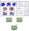
Figure 1.
Figure 1.
(A) Three-dimensional models of immunoglobulins belonging to subsets N1, N3 and N9. Antigen binding regions (panel A) and mutation analysis (panels B.1 and B.2) of modeled antibodies are shown. In all cases, Igs belonging to the same HCDR3 subset display an overall similarity, both in sequence and structure, that is not limited to the HCDR3 region. Samples 3926, 4625 and PV3 in subset N1 (panel A) have a positively charged antigen binding site (Abs) with a protruding and hydrophobic H3 loop (black arrow). Subset N3 (cases PV25 and PV52, panel A) presents nearly identical Abs. A negatively charged pocket is in the center of the binding site (black arrow). Cases PV69 and 4468 (subset N9, panel A), though presenting a less remarkable similarity, share some common features. The Abs is formed by a negatively charged central protruding region surrounded by a positive area (black arrow). Panel B.1: common mutations (i.e. replacement of the same specific residue of Igs in the same group deriving from the same germline) and convergent mutations (i.e. replacement of a residue in an Ig increasing its similarity to another Ig belonging to the same group but deriving from a different germline) are depicted in green and red, respectively, on the superimposed backbones of all the Igs in each group. Blue circled area highlights the most relevant mutations; a close-up of the same region is in panel B.2, where relevant mutations are reported in a ball-and-stick representation mapped on a single representative structure of each group. Correlated mutations are found on IGHV in subset N1, no significant correlation is evident on the light chains. Two common mutations (M33I and A71V) are located in close proximity in the models and are likely to play a role in Abs specificity. Two similar mutations (S30BN and S34N) are found in loop H1 of both samples belonging to subset N3 and may influence the H3 position and the Abs shape. Several mutations on the heavy and light chains of both samples in subset N9 introduce polar and charged residues in the Abs. Sequence numbering follows the Kabat-Chothia scheme. Abs are colored according to their electrostatic potential, ranging from -5 kBT/e (red) to + 5 kBT/e (blue). (B) Hydrophobic areas of cases belonging to subset N1. Antigen binding sites of subset N1 immunoglobulins are depicted using the Kyte-Doolittle hydropaticity score. Note the large hydrophobic patch found in the H3 loops of all samples (dark-green region) that is often associated with an interaction area.

References

    1. Swerdlow SHCE, Harris NL, Jaffe ES, Pileri SA, Stein H, Thiele J, Vardiman JW. WHO Classification of Tumours of Haematopoietic and Lymphoid Tissues. Lyon: IARC Press; 2008.
    1. Matutes E, Oscier D, Montalban C, Berger F, Callet-Bauchu E, Dogan A, et al. Splenic marginal zone lymphoma proposals for a revision of diagnostic, staging and therapeutic criteria. Leukemia. 2008;22(3):487–95. - PubMed
    1. Arcaini L, Lazzarino M, Colombo N, Burcheri S, Boveri E, Paulli M, et al. Splenic marginal zone lymphoma: a prognostic model for clinical use. Blood. 2006;107(12):4643–9. - PubMed
    1. Saadoun D, Suarez F, Lefrere F, Valensi F, Mariette X, Aouba A, et al. Splenic lymphoma with villous lymphocytes, associated with type II cryoglobulinemia and HCV infection: a new entity? Blood. 2005;105(1):74–6. - PubMed
    1. Stamatopoulos K, Belessi C, Papadaki T, Kalagiakou E, Stavroyianni N, Douka V, et al. Immunoglobulin heavy-and light-chain repertoire in splenic marginal zone lymphoma. Mol Med. 2004;10(7–12):89–95. - PMC - PubMed

Publication types