Review
. 2010 Jun;40(6):352-60.
doi: 10.2519/jospt.2010.3099.

Age-related hyperkyphosis: its causes, consequences, and management

Affiliations
Review

Age-related hyperkyphosis: its causes, consequences, and management

Wendy B Katzman et al. J Orthop Sports Phys Ther. 2010 Jun.

Abstract

Synopsis: Age-related hyperkyphosis is an exaggerated anterior curvature in the thoracic spine that occurs commonly with advanced age. This condition is associated with low bone mass, vertebral compression fractures, and degenerative disc disease, and contributes to difficulty performing activities of daily living and decline in physical performance. While there are effective treatments, currently there are no public health approaches to prevent hyperkyphosis among older adults. Our objective is to review the prevalence and natural history of hyperkyphosis, associated health implications, measurement tools, and treatments to prevent this debilitating condition.

Level of evidence: Diagnosis/prognosis/therapy, level 5.J Orthop Sports Phys Ther 2010;40(6):352-360, Epub 15 April 2010. doi:10.2519/jospt.2010.3099.


Figures
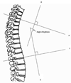
FIGURE 1
FIGURE 1
Cobb’s angle of kyphosis, calculated from a lateral radiograph. (A) Draw the first line (line a) through the superior end plate of T3, and a second line (line b) that is perpendicular to line a. (B) Draw a third line (line c) through the inferior endplate of T12, and a fourth line (line d) that is perpendicular to line c. Cobb’s angle of kyphosis is the measured angle at the intersection of lines b and d. Diagram from Kado DM, Prenovost K, Crandall C. Narrative Review: Hyperkyphosis in Older Persons. Ann Int Med. 2007;147:330–338, with permissions from Ann Int Med.
FIGURE 2
FIGURE 2
Debrunner kyphometer measurement of kyphosis. (A) Place the upper foot of the kyphometer over the interspace of T2 and T3 spinous processes, and the lower foot of the kyphometer over the interspace of T11 and T12 spinous processes. (B) Ask the patient to exhale and measure the usual kyphosis, and then “stand as straight and tall as you can” to measure the best kyphosis. (C) Read the Cobb’s angle measurement of kyphosis from the device.
FIGURE 3
FIGURE 3
Flexicurve ruler measurement of kyphosis. (A) Mark the C7 spinous process and the L5–S1 interspace on the patient’s skin with a grease pencil. (B) Place the superior end of the ruler at C7 and the inferior end over the lumbar spine, molding the ruler to the curves of the thoracic and lumbar spine. (C) Mark the level of the C7 spinous process and the L5–S1 interspace on the ruler. (D) Carefully transfer the molded ruler to tracing paper, with the C7 spinous process and the L5-S1 interspace marks aligned along a vertical line. (E) Trace the thoracic and lumbar curvatures from the ruler onto the paper, drawing a horizontal line from the vertical line to the apex of the thoracic curve. (F) Measure thoracic width (TW) and thoracic length (TL); calculate kyphosis index (KI): (TW/TL) × 100. (G) Lumbar width (LW) and lumbar length (LL) can also be measured. Photograph and diagram used with permission from Carleen Lindsey, PT, MSc, GCS, and the Section on Geriatrics, APTA.
FIGURE 4
FIGURE 4
Prone trunk lift with weighted backpack. (A) Patient lies prone over a pillow, wearing a backpack secured to the upper back. (B) Squeeze shoulder blades together, tighten gluteal muscles and lift chest off the mat, keeping cervical and lumbar spine in neutral. (C) Begin with 1 set of 10 repetitions and progress with weights in backpack, up to a maximum weight of 30% of 1-repetition maximum. In lieu of a weighted backpack, patients can use handheld dumbbells and perform the exercise with their elbows bent and their hands by their ears. Progress the dumbbells to 2.27 kg in each hand, and perform 3 sets of 8 repetitions.
FIGURE 5
FIGURE 5
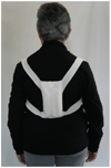
Weighted spinal kyphosis orthosis. (A) Place the weighted kyphosis orthosis over the thoracic spine and adjust the straps such that the bottom of the pouch is located at the waistline. (B) Begin with a 115-g weight in the orthosis, and progress to a 225-g weight to provide sensory feedback to improve postural alignment. (C) Instruct the patient to wear the device when ambulating.
FIGURE 6
FIGURE 6
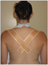
Thoracic taping for hyperkyphosis. (A) Instruct the patient to stand and elongate the crown of the head towards the ceiling. (B) Apply cover roll as needed to protect the skin. (C) Apply therapeutic tape from the anterior aspect of acromioclavicular joint, over the muscle bulk of the upper trapezius, and diagonally over the spinous process of T6. (D) Apply tape in this method bilaterally, intersecting the strips of tape at T6.

References

    1. Ball JM, Cagle P, Johnson BE, Lucasey C, Lukert BP. Spinal extension exercises prevent natural progression of kyphosis. Osteoporos Int. 2009;20:481–489. http://dx.doi.org/10.1007/s00198-008-0690-3. - DOI - PubMed
    1. Balzini L, Vannucchi L, Benvenuti F, et al. Clinical characteristics of flexed posture in elderly women. J Am Geriatr Soc. 2003;51:1419–1426. - PubMed
    1. Benedetti MG, Berti L, Presti C, Frizziero A, Giannini S. Effects of an adapted physical activity program in a group of elderly subjects with flexed posture: clinical and instrumental assessment. J Neuroeng Rehabil. 2008;5:32. http://dx.doi.org/10.1186/1743-0003-5-32. - DOI - PMC - PubMed
    1. Birnbaum K, Siebert CH, Hinkelmann J, Prescher A, Niethard FU. Correction of kyphotic deformity before and after transection of the anterior longitudinal ligament--a cadaver study. Arch Orthop Trauma Surg. 2001;121:142–147. - PubMed
    1. Bouxsein ML, Melton LJ, Riggs BL, et al. Age- and sex-specific differences in the factor of risk for vertebral fracture: a population-based study using QCT. J Bone Miner Res. 2006;21:1475–1482. - PubMed

MeSH terms