. 2010 Jul 7;29(13):2217-29.
doi: 10.1038/emboj.2010.104. Epub 2010 May 28.

A novel ATP-dependent conformation in p97 N-D1 fragment revealed by crystal structures of disease-related mutants

Affiliations

A novel ATP-dependent conformation in p97 N-D1 fragment revealed by crystal structures of disease-related mutants

Wai Kwan Tang et al. EMBO J. .

Abstract

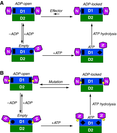
Mutations in p97, a major cytosolic AAA (ATPases associated with a variety of cellular activities) chaperone, cause inclusion body myopathy associated with Paget's disease of the bone and frontotemporal dementia (IBMPFD). IBMPFD mutants have single amino-acid substitutions at the interface between the N-terminal domain (N-domain) and the adjacent AAA domain (D1), resulting in a reduced affinity for ADP. The structures of p97 N-D1 fragments bearing IBMPFD mutations adopt an atypical N-domain conformation in the presence of Mg(2+).ATPgammaS, which is reversible by ADP, showing for the first time the nucleotide-dependent conformational change of the N-domain. The transition from the ADP- to the ATPgammaS-bound state is accompanied by a loop-to-helix conversion in the N-D1 linker and by an apparent re-ordering in the N-terminal region of p97. X-ray scattering experiments suggest that wild-type p97 subunits undergo a similar nucleotide-dependent N-domain conformational change. We propose that IBMPFD mutations alter the timing of the transition between nucleotide states by destabilizing the ADP-bound form and consequently interfere with the interactions between the N-domains and their substrates.


Conflict of interest statement

The authors declare that they have no conflict of interest.

Figures

Figure 1
Figure 1
Structures of wild-type and mutant p97 N–D1 with bound ADP or ATPγS. Mutations, either identified in IBMPFD patients (yellow) or introduced in this work (R86A in red and R53A in green), are shown as ball-and-stick models. The N-domains are in magenta and the colours of the D1-domains are alternating in grey (cognate of N) and blue. (A) A schematic diagram for the domain arrangement of p97. (B) Mapping of various mutations to the Cα trace of the wild-type p97 hexamer with bound ADP (PDB:1E32) (Zhang et al, 2000). The structure is viewed down the six-fold axis from the D1- to D2-domain. (C) Stereoscopic rendition of a magnified portion in Figure 1B showing a detailed distribution of mutations in p97 with bound ADP. All mutation sites are labelled. (D) Mapping of various mutations to the Cα trace of the mutant (R155H) structure with bound Mg2+·ATPγS. (E) A stereo pair showing a detailed distribution of the mutation sites in the ATPγS-bound structure. (F) Superposition of the hexamers of the ATPγS and ADP forms. The two structures are portrayed in Cα traces with the N-domain from the ATPγS form in pink and that from the ADP form in magenta. Diameters and heights for the hexamers are given. (G) A stereo pair showing a portion of the superposition in Figure 1F. The superposition was based solely on the D1 RecA-like domain. The ADP form shows the N-domain in magenta, N–D1 linker in forest green, and D1-domain in dark blue, whereas the ATPγS form has its N-domain coloured in pink, N–D1 linker in light green, and D1-domain in light blue.
Figure 2
Figure 2
Nucleotide-binding-associated conformational changes in solution observed by X-ray scattering experiments. (A) Guinier plots (Guinier and Fournet, 1955) for the wild-type and mutant p97 N–D1 fragments in the presence of 0.1 mM ATPγS or ADP. Wild-type data are in red, R95G and R155H mutants are in black and green, respectively. Open circles are for ADP and open triangles for ATPγS. (B) Distance distribution functions, p(r), normalized to a common total probability, with standard error of the data in Å propagated through the inverse Fourier transform implemented in GNOM (Svergun, 1992) for the wild-type and mutant N–D1 fragments. The calculated distribution (Glatter, 1980) is given in the last panel based on crystal structure in the absence of bound-solvent molecules. The dashed and solid lines are for ATPγS and ADP, respectively.
Figure 3
Figure 3
Changes in the structure of N–D1 on binding of ATPγS. (A) Stereoscopic pair showing details of the ATPγS-binding environment. The nucleotide-binding site is located at the subunit interface. One subunit is coloured in green and the other in grey. Residues making contact with bound ATPγS are drawn as sticks and are labelled. The ATPγS molecule is shown as a stick model with carbon in purple, oxygen in red, nitrogen in blue, phosphorous in magenta, and sulphur in yellow. The ATPγS molecule is enclosed in a difference electron density cage in grey, contoured at the 2.5σ level. The Mg2+ ion is shown as a green ball with the three coordinating water molecules in red. (B) Stereo pair showing the ordering of an N-terminal segment from Leu12 to Lys20 in the ATPγS form and its environment. The re-ordered residues are depicted as stick models in yellow and their environment is shown as a semi-transparent molecular surface with all interacting residues rendered as stick models with labels. The yellow surface is the N-domain; orange and green surfaces are the cognate D1-domain and N–D1 linker, respectively. The grey surface is from an adjacent D1-domain. (C) Conformation of the N–D1 linker in the presence of ATPγS. The N–D1 linker is shown as a ribbon model in green; N- and D1-domains are shown as molecular surfaces in magenta and in blue, respectively. The bound ATPγS is shown as a stick model. (D) Conformation of the N–D1 linker in the presence of ADP. The N–D1 linker is shown as a coil in green. The D1-domain is similarly orientated as in (C). The bound ADP is shown as a stick model.
Figure 4
Figure 4
Calorimetric titration of wild-type and mutant p97 N–D1 with ATPγS or ADP. (A) ITC for wild-type p97 with ADP. Raw data was obtained from 27 automatic injections of 10 μl of 100 μM ADP solution (first injection 2 μl) into 1.4236 ml of 10 μM p97 solution at 25°C (top panel) and fitted (bottom panel, continuous line) to a one-site model. (B) ITC for wild-type p97 with ATPγS; 30 injections of 8 μl of 100 μM ATPγS solution were made into 20 μM p97 solution (top panel). The data was fitted to a one-site model (bottom panel). (C) ITC for the R95G mutant with ADP; 27 injections of 10 μl of 100 μM ADP solution were made into 10 μM mutant p97 solution (top panel). The resulting heat data was fitted to a one-site model (bottom panel). (D) ITC for the R95G mutant with ATPγS; 30 injections of 10 μl of 100 μM ATPγS solution were made into 10 μM mutant p97 solution (top panel). The data was fitted to a two-site model (bottom panel).
Figure 5
Figure 5
Mechanistic model for the control of N-domain movement and implication for IBMPFD. Schematic diagram for the control of the N-domain conformation in (A) the wild-type and (B) IBMPFD mutant p97 N–D1 fragment. The N-, D1-, and D2-domains are in magenta, blue, and green, respectively, and as labelled. The small yellow circles between N- and D1-domains represent positions of mutations. Four states are defined for each nucleotide-binding site in D1: Empty state, ATP state, ADP-locked, and ADP-open, as labelled. Each protomer is assumed to operate independently. The stimuli for changes in D1 nucleotide state may come either from the N-domain or from ATP hydrolysis of the D2-domain. In the ADP-locked state, the N-domains are in Down-conformation with a pre-bound ADP shown as a black rectangle. This ADP-locked state has been observed crystallographically in wild-type p97. In the ATP state, the N-domains of hexameric wild-type p97 could be either in an Up-conformation with bound ATP shown as a black diamond or in a Down-conformation in an ADP-locked state, whereas the N-domains of mutants adopt only the Up-conformation as observed in this study. On the basis of available structural and biochemical information, we introduce two additional conformational states. The Empty state has the N-domain conformation undefined and the nucleotide-binding site shown as a black circle. The ADP-open state also has an N-domain conformation similar to that of Down-conformation as determined by the crystal structure of R155H mutant with bound ADP, which is also shown as a black rectangle. Bound ADP can only be exchanged through the ADP-open state. In wild-type p97, the equilibration between ADP-open and ADP-locked favours heavily the latter and is presumably regulated by effectors such as p47 or ATP hydrolysis in D2-domain. In IBMPFD mutants, the tight control between ADP-open and ADP-locked is disrupted and the equilibration is now favouring the ADP-open state.

Similar articles

Cited by

References

    1. Beuron F, Dreveny I, Yuan X, Pye VE, McKeown C, Briggs LC, Cliff MJ, Kaneko Y, Wallis R, Isaacson RL, Ladbury JE, Matthews SJ, Kondo H, Zhang X, Freemont PS (2006) Conformational changes in the AAA ATPase p97-p47 adaptor complex. EMBO J 25: 1967–1976 - PMC - PubMed
    1. Beuron F, Flynn TC, Ma J, Kondo H, Zhang X, Freemont PS (2003) Motions and negative cooperativity between p97 domains revealed by cryo-electron microscopy and quantised elastic deformational model. J Mol Biol 327: 619–629 - PubMed
    1. Briggs LC, Baldwin GS, Miyata N, Kondo H, Zhang X, Freemont PS (2008) Analysis of nucleotide binding to P97 reveals the properties of a tandem AAA hexameric ATPase. J Biol Chem 283: 13745–13752 - PMC - PubMed
    1. Dai RM, Li CC (2001) Valosin-containg protein is a multi-ubiquitin chain-targeting factor required in ubiquitin-proteasome degradation. Nat Cell Biol 3: 740–744 - PubMed
    1. Davies JM, Tsuruta H, May AP, Weis WI (2005) Conformational changes of p97 during nucleotide hydrolysis determined by small-angle X-ray scattering. Structure 13: 183–195 - PubMed

Publication types

MeSH terms