

doi: 10.1038/nbt.1631.
Epub 2010 May 30.
Synthetic polymer coatings for long-term growth of human embryonic stem cells
Affiliations
- PMID: 20512122
- PMCID: PMC3471651
- DOI: 10.1038/nbt.1631
Item in Clipboard
Synthetic polymer coatings for long-term growth of human embryonic stem cells
Nat Biotechnol.
2010 Jun.
Abstract
We report a fully defined synthetic polymer coating, poly[2-(methacryloyloxy)ethyl dimethyl-(3-sulfopropyl)ammonium hydroxide] (PMEDSAH), which sustains long-term human embryonic stem (hES) cell growth in several different culture media, including commercially available defined media. The development of a standardized, controllable and sustainable culture matrix for hES cells is an essential step in elucidating mechanisms that control hES cell behavior and in optimizing conditions for biomedical applications of hES cells.
Figures
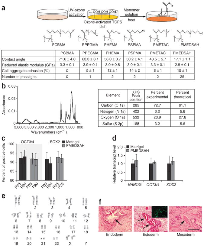
Long-term culture of H9 hES cells on PMEDSAH with MEF-conditioned media. (a) Schematic diagram showing graft-polymerization used to synthesize the polymer coatings and their chemical structures. Tissue culture polystyrene dishes were first activated by UV ozone and then methacrylate-based monomers were polymerized from the surface. Table lists contact angle, reduced elastic modulus (GPa) (mean ± s.d.), initial hES cel aggregate adhesion (mean ± standard error) and number of passages achieved on each polymer coating. (b) Fourier transform infrared spectrum of PMEDSAH coating showing distinct bands at 1,732.9 cm−1 and 1,208.4 cm−1, which indicated presence of carbonyl and sulfonate groups, respectively. Table lists elemental composition of PMEDSAH obtained using X-ray photoelectron spectroscopy. Relative composition of these elements was in agreement with the expected chemical composition of PMEDSAH. (c) Percentage (mean ± standard error) of hES cells expressing OCT3/4 and SOX2 at passages 3 (P03) and 20 (P20). (d) Relative transcript levels of NANOG, OCT3/4 and SOX2 from hES cells cultured on PMEDSAH and Matrigel. (e,f) After 25 passages, hES cells cultured on PMEDSAH and Matrigel (Supplementary Fig. 3) (e) maintained a normal karyotype and (f) retained pluripotency as demonstrated by teratoma formation in mmunosuppressed mice. H&E-stained paraffn sections indicating endoderm (goblet-like cells at arrow), ectoderm (neuroepithelial aggregates at arrow; and cells expressing neuron-restricted protein β-III tubulin in inset) and mesodermal derivatives (cartilage, connective tissue and muscle at arrow). Scale bar, 200 μm.
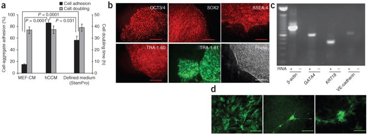
PMEDSAH supports culture of hES cells in defined medium. (a) Percentage (mean ± standard error) of cell aggregate adhesion (number of aggregates attached with respect to total aggregates passaged) and population doubling time (twofold increase in colony area) for H9 hES cells cultured on PMEDSAH in MEF-conditioned medium, human cell–conditioned medium and defned medium. P-values calculated using unpaired t-test. (b) Fluorescence micrographs of colonies of H9 cells cultured on PMEDSAH in StemPro medium showing expression of hES cell markers: OCT3/4, SOX2, SSEA-4, TRA-1-60 and TRA-1-81; and a phase-contrast image. (c) RT-PCR analysis of RNA from embryoid bodies showing expression of endoderm (GATA4), ectoderm (KRT18) and mesoderm derivatives (VE-cadherin; also known as CDH5). β-Actin (also known as ACTB) was used as positive control, and for each primer set tested, a reaction lacking RNA was assessed in parallel as a negative control. Scale bars, 200 μm, except for SOX2, which is 100 μm (d) Micrographs showing immunoreactivity for α-fetoprotein (endoderm), β-III tubulin (ectoderm) and smooth muscle actin (mesoderm) demonstrating the pluripotent state of H9 cells cultured on PMEDSAH in StemPro medium. Scale bars, 200 μm.
Comment in
-
Scalable pluripotent stem cell culture.Nat Biotechnol. 2010 Jun;28(6):562-3. doi: 10.1038/nbt0610-562. Nat Biotechnol. 2010. PMID: 20531334 No abstract available.
-
Matrix revolutions: a trinity of defined substrates for long-term expansion of human ESCs.Cell Stem Cell. 2010 Jul 2;7(1):7-8. doi: 10.1016/j.stem.2010.06.008. Cell Stem Cell. 2010. PMID: 20621041
References
Publication types
MeSH terms
Substances
Grants and funding
LinkOut - more resources
Full Text Sources
Other Literature Sources
