. 2010 Jul;13(7):861-8.
doi: 10.1038/nn.2581. Epub 2010 May 30.

Specific roles for DEG/ENaC and TRP channels in touch and thermosensation in C. elegans nociceptors

Affiliations

Specific roles for DEG/ENaC and TRP channels in touch and thermosensation in C. elegans nociceptors

Marios Chatzigeorgiou et al. Nat Neurosci. 2010 Jul.

Abstract

Polymodal nociceptors detect noxious stimuli, including harsh touch, toxic chemicals and extremes of heat and cold. The molecular mechanisms by which nociceptors are able to sense multiple qualitatively distinct stimuli are not well understood. We found that the C. elegans PVD neurons are mulitidendritic nociceptors that respond to harsh touch and cold temperatures. The harsh touch modality specifically required the DEG/ENaC proteins MEC-10 and DEGT-1, which represent putative components of a harsh touch mechanotransduction complex. In contrast, responses to cold required the TRPA-1 channel and were MEC-10 and DEGT-1 independent. Heterologous expression of C. elegans TRPA-1 conferred cold responsiveness to other C. elegans neurons and to mammalian cells, indicating that TRPA-1 is a cold sensor. Our results suggest that C. elegans nociceptors respond to thermal and mechanical stimuli using distinct sets of molecules and identify DEG/ENaC channels as potential receptors for mechanical pain.


Figures

Figure 1
Figure 1. PVD neurons respond to harsh touch and cold temperature.
a–c. PVD neurons display complex dendritic arbors that envelop the animal. Confocal Z-projection of adult (left side) showing PVDL labeled with F49H12.4::GFP marker (anterior to left, ventral side corresponds to inside surface of looped worm). PVDR on the right side is excluded from this confocal series. Insets show enlarged view of posterior-lateral location of PVDL cell soma with ventrally projecting axon and orthogonal array of dendritic branches. Scale bar in panel A is 15 μm. d–e. Calcium responses to harsh (d) and gentle (e) touch. Each red trace represents the average percentage change in normalized YFP/CFP ratio (R/R0) for 20 individual recordings. The black triangle indicates the time at which the mechanical stimulus (see Methods) was applied. Gray shading indicates SEM of the mean response. f. Behavioral responses to cold shock. Shown are percentages of animals (n=30) displaying avoidance behavior (omega bends) during a recording of animals experiencing acute temperature change (20-15° C). The blue box indicates a 50 second interval during which the buffer temperature was 15° C; temperature was 20° C during the remainder of the recording. Error bars indicate SEM; PVD- animals (in which both PVD neurons ablated by laser microsurgery) were significantly less responsive than intact animals (* P< .05) according to the Student’s t test. g. Calcium response to cold shock. Red trace represents the average percentage change in R/R0 for 20 individual recordings; gray shading indicates SEM of the mean response. The lower line indicates the buffer temperature during the recording.
Figure 2
Figure 2. mec-10 is required for harsh touch in PVD
a–b mec-10(tm1552) animals are defective in harsh touch response in PVD. Shown are averaged responses of 17 wild-type (a), and 14 mec-10(tm1552) mutant (b) animals to harsh body touch. For these and other panels, the red trace represents the average percentage change in normalized YFP/CFP ratio (R/R0); gray shading indicates SEM of the mean response. Scale bars are indicated in upper left. The triangle indicates the time of the stimulus. c. PVD-specific rescue of the mec-10 harsh touch phenotype. Shown is the averaged calcium response of 13 mec-10(tm1552); ljEx221 [pser-2prom-3::mec-10(+)] animals. Rescue was also observed with a mec-10 cDNA and with a second PVD-specific promoter, pegl-46 (see panel F). d–e. trpa-1 and osm-9 are not required for harsh touch in PVD. Shown are averaged traces for 13 trpa-1(ok999) (D) and 17 osm-9(ky10) (E) animals. f. Scatter plot of peak calcium responses for each genotype. Statistical significance (*** p < .0005) is according to the Mann-Whitney rank sum test. Additional genotypes shown include mec-10(tm1552); ljEx220 [pegl-46::mec-10(+)] (13 recordings) and mec-10(tm1552); ljEx230 [pser-2prom-3::mec-10(cDNA)] (12 recordings).
Figure 3
Figure 3. DEGT-1 is required for harsh touch responses in PVD
a–c. Calcium responses of wild-type and degt-1RNAi animals to harsh body touch in PVD. Each red trace represents the average percentage change in normalized YFP/CFP ratio (R/R0) for the indicated genotype; gray shading indicates SEM of the mean response. Panel a shows wild-type response (13 animals recorded). Panels b and c show loss of harsh touch response in lines expressing degt-1 RNAi under the PVD-specific promoters pegl-46 (ljEx224; 14 animals) and pser-2prom3 (ljEx225; 17 animals). In panel c, the green line shows rescue of the pser-2::degt-1RNAi (ljEx225) phenotype by the C. briggsae orthologue Cbr-degt-1 expressed cell-specifically in PVD under the control of the pser-2prom3 promoter (ljEx261; 14 animals). Cbr-degt-1 shares less than 5% sequence identity with C. elegans degt-1 over the region targeted by ljEx225. d. Scatter plot of peak calcium responses for each genotype. Statistical significance (*** p < .001) is according to the Mann-Whitney rank sum test. Also shown are responses of the off-target control in which a degt-1 RNAi transgene is expressed outside PVD under the touch neuron promoter pmec-4 (ljEx240; 14 animals recorded). Responses of additional on-target and off-target RNAi control strains are shown in Supplemental Figure 4.
Figure 4
Figure 4. Localization patterns of DEGT-1 and MEC-10 fusion proteins in PVD
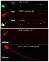
For all figures, the large arrows indicate the PVD soma. a–c. Colocalization of MEC-10::GFP and DEGT-1::RFP in PVD dendritic puncta. Shown are green, red, and dual wavelength images of a single confocal section of AQ2427 ljEx250[pser-2prom3::mec-10::GFP; pmyo-2::GFP]; ljEx256[pser-2prom3::degt-1::mCherry pmyo-2::GFP] animals, which express both MEC-10::GFP and DEGT-1::RFP in PVD. AQ2427 was generated by crossing AQ2396 ljEx250 and AQ2402 lj256, and selecting for the presence of both arrays. These images all show the primary PVD dendrite; similar results were observed in higher-order dendritic branches (Supplemental Figure 6). d–e. Punctate localization of DEGT-1::RFP is mec-10-dependent. Shown are single confocal sections of PVD processes expressing the ljEx256 DEGT-1::RFP transgene in a wild-type (d) or mec-10(tm1552) mutant (e) background. degt-1 RNAi did not abolish punctate expression of MEC-4::GFP (Supplemental Figure 6).
Figure 5
Figure 5. Effect of mec-10 on harsh touch responses in ALM
a–b. mec-10 is required for mec-4-independent harsh touch response in ALM. Shown are calcium responses in ALM to fast large-displacement stimulation in mec-4(u253) single mutant (a) and mec-10(tm15520 mec-4(u253) double mutant (b). In these and other panels, each red trace represents the average percentage change in R/R0; gray shading indicates SEM of the mean response. Triangle indicates application of the harsh touch stimulus. Panel a represents the averaged response of 13 mec-4(u253) animals; panel b represents the response of 13 mec-10(tm1552) mec-4(u253) animals. c. mec-10 functions in the body touch neurons to promote ALM harsh touch response. Shown is the averaged response of 17 mec-10(tm1552) mec-4(u253); pmec-4::mec-10(+) animals, in which mec-10 is specifically rescued in the body touch neurons. Similar results were seen when the PVD neurons were eliminated by laser ablation (Supplemental Figure 7). d. degt-1 is required for ALM harsh touch responses. Shown is the averaged response of 19 mec-4(u253); ljEx240[pmec-4::degt-1RNAi] animals, in which degt-1 is specifically eliminated in the ALM neurons by RNAi. Additional off-target RNAi controls are shown in Supplemental Figure 4C. e. Scatter plot of peak calcium responses for each genotype. Statistical significance (*** p < .001;) is according to the Mann-Whitney rank sum test.
Figure 6
Figure 6. TRPA-1 is specifically required for cold responses in PVD
a. trpa-1 is required for PVD cold response. Red trace indicates the averaged percentage change in R/R0 of 17 trpa-1(ok999) animals; green trace is the response of 13 trpa-1(ok999)); ljEx245 [pegl-46::trpa-1(+)] animals in which a wild-type trpa-1(+) transgene cell-specifically rescues the null phenotype in PVD. Lower line indicates temperature changes during the recording; gray shading indicates SEM of the mean response. Scale bars are indicated in upper left. b. trpa-1 is required in PVD for cold shock avoidance behavior. Shown are percentages of trpa-1(ok999) and PVD-rescued animals displaying avoidance behavior (omega turns) following acute temperature change (20-15° C). Blue boxes indicate the duration of the 15° C cold shock; error bars indicate SEM. c. Mammalian mTRPA1 confers cold responses in PVD. Red trace indicates the averaged response of 13 trpa-1(ok999)); ljEx262 [pegl-46::mTRPA1] animals. d. Scatter plot of peak calcium responses for each genotype. Statistical significance (*** p < .001) is according to the Mann-Whitney rank sum test. e–f. Subcellular localization of a rescuing full-length TRPA-1::GFP fusion in PVD. Fluorescence images of AQ2428 ljEx267[pser-2prom3::trpa-1::GFP pmyo-2::GFP], which express a rescuing full-length TRPA-1::GFP fusion protein in PVD. Panel e shows a single confocal section with DIC optics, while panel f shows a Z-stack confocal projection in darkfield. Phenotypic rescue of the trpa-1 cold avoidance phenotype is shown in Supplemental Figure 5.
Figure 7
Figure 7. Heterologous expression of TRPA-1 in C. elegans neurons confers cold sensitivity
a. Normal response of FLP to noxious heat shock. Averaged calcium trace of wild-type FLP neurons expressing the ljEx19 transgene. For this and other panels, the red trace represents the average percentage change in R/R0 for 20 individual animals. Gray shading indicates SEM of the mean response, lower line indicates temperature changes during the recording. Scale bars are indicated in upper right. b. FLP does not respond to cold shock in wild type animals. Shown is an averaged trace of 20 wild-type ljEx19 animals in response to 20°-15° C cold shock. c. Animals expressing trpa-1(+) ectopically in FLP respond to cold shock. Trace shows averaged response of 20 trpa-1(ok999); ljEx246[pegl-46::trpa-1(+)]; ljEx19 animals, which express trpa-1(+) heterologously in FLP. d. Wild-type ALM neurons do not respond to cold. Shown is the averaged response of 20 bzIs17[pmec-4::YC2.12] animals, which express cameleon in the body touch neurons. e. Animals expressing trpa-1(+) ectopically in ALM respond to cold shock. Shown is the averaged response of 20 trpa-1(ok999); ljEx223 [pmec-4::trpa-1(+)]; bzIs17 animals, in which trpa-1(+) is expressed heterologously in ALM and other the body touch neurons. f. Scatter plot of peak calcium responses for each genotype. Statistical significance (*** p < .001) is according to the Mann-Whitney rank sum test.
Figure 8
Figure 8. Cold stimuli activate TRPA-1-expressing HEK293T cells
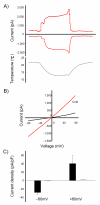
a. TRPA-1-expressing HEK cells respond to cold in whole-cell configuration. Perfusion of the cold bath solution activates a representative TRPA-1-expressing HEK cell (n=9). Currents are shown at holding potentials of +60 and −60 mV. As previously observed in CHO cells , currents were also activated by pressure (Supplemental Figure 11). b. Instantaneous current voltage relationships of the TRPA-1-expressing HEK cell are shown. Voltages were ramped from −80 to +80 mV. Responses are shown before and during application of 15°C cold temperature in the bath. c. Average current densities evoked by a cold temperature of 15°C. Current densities of the cold responses of TRPA-1-transfected (filled bars) and untransfected HEK cells (open bars) at ± 60 mV (n=9 for cold response, n=26 for no response). Error bars, ± SEM.

Similar articles

Cited by

References

    1. Patapoutian A, Tate S, Woolf CJ. Transient receptor potential channels: targeting pain at the source. Nat Rev Drug Discov. 2009;8:55–68. - PMC - PubMed
    1. Wemmie JA, Price MP, Welsh MJ. Acid-sensing ion channels: advances, questions and therapeutic opportunities. Trends Neurosci. 2006;29:578–86. - PubMed
    1. Caterina MJ, et al. The capsaicin receptor: a heat-activated ion channel in the pain pathway. Nature. 1997;389:816–24. - PubMed
    1. Tominaga M, et al. The cloned capsaicin receptor integrates multiple pain-producing stimuli. Neuron. 1998;21:531–43. - PubMed
    1. McKemy DD, Neuhausser WM, Julius D. Identification of a cold receptor reveals a general role for TRP channels in thermosensation. Nature. 2002;416:52–8. - PubMed

Publication types

MeSH terms

Substances