
Mutations in TMEM216 perturb ciliogenesis and cause Joubert, Meckel and related syndromes

Enza Maria Valente et al. Nat Genet. 2010 Jul.

Abstract

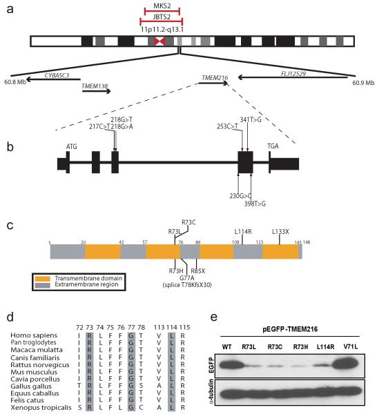
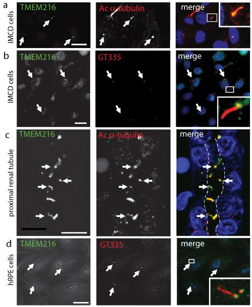
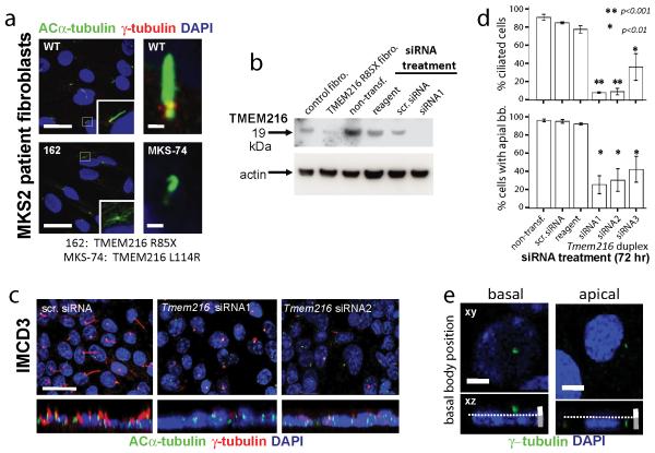
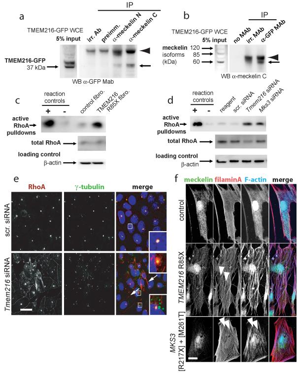
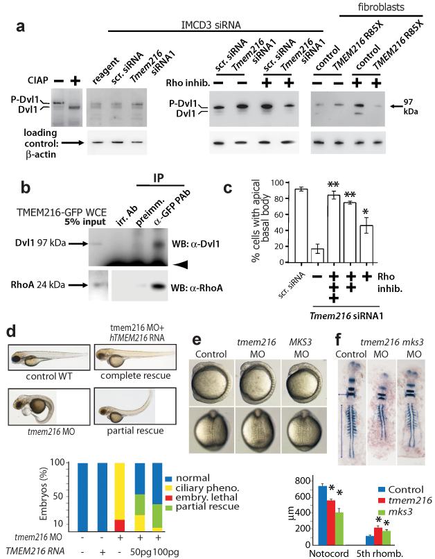
Joubert syndrome (JBTS), related disorders (JSRDs) and Meckel syndrome (MKS) are ciliopathies. We now report that MKS2 and CORS2 (JBTS2) loci are allelic and caused by mutations in TMEM216, which encodes an uncharacterized tetraspan transmembrane protein. Individuals with CORS2 frequently had nephronophthisis and polydactyly, and two affected individuals conformed to the oro-facio-digital type VI phenotype, whereas skeletal dysplasia was common in fetuses affected by MKS. A single G218T mutation (R73L in the protein) was identified in all cases of Ashkenazi Jewish descent (n=10). TMEM216 localized to the base of primary cilia, and loss of TMEM216 in mutant fibroblasts or after knockdown caused defective ciliogenesis and centrosomal docking, with concomitant hyperactivation of RhoA and Dishevelled. TMEM216 formed a complex with Meckelin, which is encoded by a gene also mutated in JSRDs and MKS. Disruption of tmem216 expression in zebrafish caused gastrulation defects similar to those in other ciliary morphants. These data implicate a new family of proteins in the ciliopathies and further support allelism between ciliopathy disorders.


Figures

Figure 1
Figure 1
Mutations in the TMEM216 gene in patients linked to the JBTS2 and MKS2 loci. (a) Chromosomal location of the JBTS2 and MKS2 loci on Chr. 11cent. (b) TMEM216 genomic organization, depicting start and stop codon, and location of identified base changes. (c) The longest splice isoform encodes for a 148 aa tetraspan membrane protein. Patient mutations predominate towards the middle, with one prevalent p.R73 change occurring repeatedly. Missense, nonsense and splice mutations were identified. (d) Evolutionary conservation of mutated amino acids. (e) Patient mutations lead to unstable protein products. Western blot of whole lysate of cells transfected with a cDNA encoding wild type (WT) vs. patient missense mutations, compared with control (p.V71L). Each mutation resulted in the production of 40-50% of WT protein levels, compared with α-tubulin loading control.
Figure 2
Figure 2
Ciliary localization of TMEM216. (a-d) Overlapping localization of endogenous TMEM216 (green) and acetylated α-tubulin or GT335 (glutamylated tubulin) (red) at the base of the primary cilium (arrows) in IMCD3 (a, b), proximal renal tubules (c) or hRPE cells (d). White dashed line indicates the tubule lumen. Boxes show insets at magnification x10. Scale bar 5 μm.
Figure 3
Figure 3
TMEM216 mutation or knockdown results in impaired ciliogenesis and centrosome docking. (a) Two different TMEM216-mutated patient fibroblasts lines show defective ciliogenesis and impaired centrosome docking (marked by γ-tubulin). Scale bar: left 20 μm; right 1 μm. (b) TMEM216 antisera reacts with a 19 kDa band in control cells, which is reduced in TMEM216 p.R85X fibroblasts (some residual is apparent likely due to read-through from geneticin treatment), as well as in siRNA1-treated IMCD3 cells. Fibro. = fibroblasts; Non-transf. = non-transfected; scr. = scrambled. (c) Transfected IMCD3 cells showing effect of Tmem216 siRNA treatment, with reduced ciliogenesis and centrosome docking (note lack of cilia and lack of apically located centrosomes following knockdown). Top is x-y, and bottom is x-z projection, scale bar 10 μm. (d) Percent ciliated cells (defined as cilia > 1 μm length) is reduced following Tmem216 siRNA treatment. Percent cells with apical basal bodies (defined as most superior 1.0 μm sections compared to nuclear position) is similarly reduced. *p<0.01, **p<0.001, chi-squared test. (e) Shows method of quantification at 72 hrs. Scale bars: white, most apical 1.0 μm; grey, basal 1.5 μm.
Figure 4
Figure 4
TMEM216 complexes with Meckelin, and their loss results in Rho hyperactivation and actin cytoskeleton remodelling. (a) TMEM216-GFP (~37 kDa; arrow) is immunoprecipitated (IP) with anti-Meckelin antisera against either the N- or C-termini from whole cell extract (input WCE), but not in control IPs with an irrelevant antibody (irr. Ab) or the preimmune antiserum (preimm.). Arrowhead is IgG heavy chain. (b) IP of TMEM216-GFP by α-GFP pulls down a 60 kDa C-terminal containing isoform of endogenous Meckelin (arrow), but not in control IPs with no antibody (no MAb) or an irrelevant antibody (irr. MAb). Arrowhead is IgG heavy chain. (c) MKS2 fibroblast (fibro.) WCE has increased levels of activated RhoA-GTP compared to normal control. (d) siRNA knockdown of Tmem216 and Mks3 in IMCD3 cells increased RhoA activation, compared with scrambled control (scr.). Total RhoA and β-actin are loading controls. Positive control (+) is loading with non-hydrolyzable GTPγS, negative control (−) is loading with GDP. (e) RhoA (red) localizes to the basal bodies (γ-tubulin, green) in IMCD3 cells following 24 hr treatment with scrambled siRNA, but mislocalizes to regions adjacent to the basal bodies (arrows; and inset, magnification x5) and at basolateral surfaces (arrowheads) following Tmem216 knockdown. Mislocalization of γ-tubulin is also apparent (bottom inset). Scale bar 10 μm. (f) Subcellular phenotypes of fibroblasts cultured from undiseased control and two MKS fetuses mutated in TMEM216 [p.R85X homozygous] and MKS3 [p.R217X]+[p.M261T], as indicated. Actin stress fibers in both mutated cells (arrowheads) are detected by phalloidin staining. Scale bar 10 μm.
Figure 5
Figure 5
TMEM216 disruption results in Dvl1 phosphorylation, and planar cell polarity-like phenotypes in zebrafish. (a) siRNA knockdown of Tmem216 (left panel) and TMEM216 p.R85X patient fibroblasts (right panel) cause an increase in the upper (phosphorylated) isoform (P-Dvl1) compared to scrambled control (scr.). Treatment with Rho inhibitor exoenzyme-C3-transferase (2 μg/ml) alone increased Dvl1 phosphorylation, but increases in P-Dvl1 by TMEM216 loss are reversed by Rho inhibition (right panel). (b) Co-immunoprecipitation of both Dvl1 and RhoA with TMEM216 in TMEM216-GFP transfected cells. Arrowhead is IgG heavy chain. (c) Dose-dependent rescue of centrosome/basal body docking phenotype in Tmem216 siRNA-treated cells following += 0.5, ++= 1.0, +++= 2.0 μg/ml Rho inhibitor treatment. *p<0.01; **p<0.001 for chi-squared test. (d) Injection of translation-blocking morpholino (MO) to tmem216 vs. scrambled MO causes a ciliary defect phenotype in injected zebrafish embryos (>50 each condition). Injection of human TMEM216 RNA causes no phenotype in WT embryos, but allows partial, dose-dependent rescue of the MO phenotype. (e) Lateral (top) and dorsal (bottom) views of zebrafish embryos injected with tmem216 or mks3 MO at 8-somite stage had ciliopathy features. (f) Representative 11-somite stage embryos hybridized with krox20, pax2, and myoD riboprobes. Convergence to the midline is measured by the width at the fifth rhombomere (horizontal arrow), and extension along the anterior-posterior (AP) axis by notochord length (vertical arrow) (n=12-15 embryos/injection). Suppression of the tmem216 or mks3 morphant gastrulation defect causes significant differences in width and length compared to controls (*p<0.005). Pheno.=phenotype; embry.=embryonic; rhomb.=rhombomere; Bars=standard error of means.

Comment in

Similar articles

Cited by

References

    1. Lancaster MA, Gleeson JG. The primary cilium as a cellular signaling center: lessons from disease. Curr Opin Genet Dev. 2009;19:220–9. - PMC - PubMed
    1. Keeler LC, et al. Linkage analysis in families with Joubert syndrome plus oculo-renal involvement identifies the CORS2 locus on chromosome 11p12-q13.3. Am J Hum Genet. 2003;73:656–62. - PMC - PubMed
    1. Valente EM, et al. Description, nomenclature, and mapping of a novel cerebello-renal syndrome with the molar tooth malformation. Am J Hum Genet. 2003;73:663–70. - PMC - PubMed
    1. Valente EM, et al. Distinguishing the four genetic causes of Joubert syndrome-related disorders. Ann Neurol. 2005;57:513–9. - PubMed
    1. Baala L, et al. The Meckel-Gruber syndrome gene, MKS3, is mutated in Joubert syndrome. Am J Hum Genet. 2007;80:186–94. - PMC - PubMed

Publication types

MeSH terms