. 2010 Oct;18(10):1100-6.
doi: 10.1038/ejhg.2010.79. Epub 2010 May 26.

New RAB3GAP1 mutations in patients with Warburg Micro Syndrome from different ethnic backgrounds and a possible founder effect in the Danish

Affiliations

New RAB3GAP1 mutations in patients with Warburg Micro Syndrome from different ethnic backgrounds and a possible founder effect in the Danish

Deborah J Morris-Rosendahl et al. Eur J Hum Genet. 2010 Oct.

Abstract

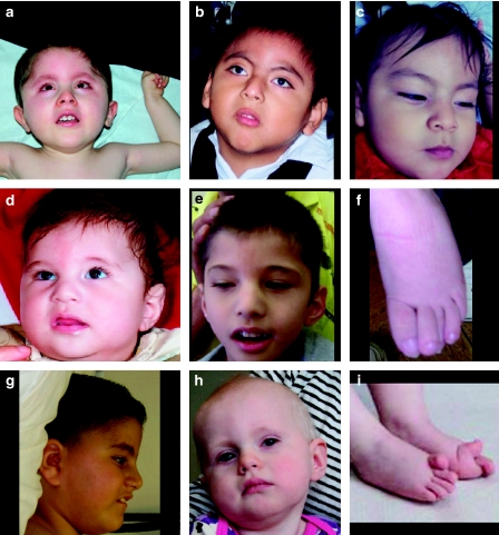
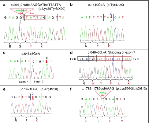
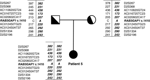
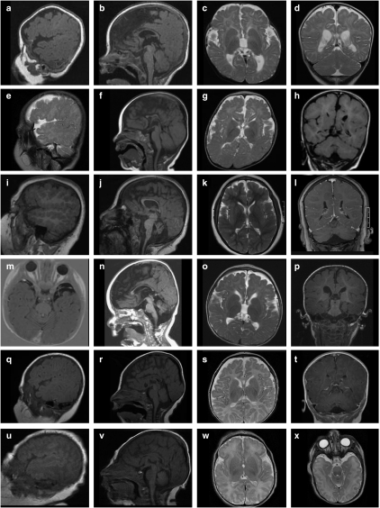
Warburg Micro Syndrome is a rare, autosomal recessive syndrome characterized by microcephaly, microphthalmia, microcornia, congenital cataracts, optic atrophy, cortical dysplasia, in particular corpus callosum hypoplasia, severe mental retardation, spastic diplegia, and hypogonadism. We have found five new mutations in the RAB3GAP1 gene in seven patients with suspected Micro Syndrome from families with Turkish, Palestinian, Danish, and Guatemalan backgrounds. A thorough clinical investigation of the patients has allowed the delineation of symptoms that are consistently present in the patients and may aid the differential diagnosis of Micro Syndrome for patients in the future. All patients had postnatal microcephaly, micropthalmia, microcornia, bilateral congenital cataracts, short palpebral fissures, optic atrophy, severe mental retardation, and congenital hypotonia with subsequent spasticity. Only one patient had microcephaly at birth, highlighting the fact that congenital microcephaly is not a consistent feature of Micro syndrome. Analysis of the brain magnetic resonance imagings (MRIs) revealed a consistent pattern of polymicrogyria in the frontal and parietal lobes, wide sylvian fissures, a thin hypoplastic corpus callosum, and increased subdural spaces. All patients were homozygous for the mutations detected and all mutations were predicted to result in a truncated RAB3GAP1 protein. The analysis of nine polymorphic markers flanking the RAB3GAP1 gene showed that the mutation c.1410C>A (p.Tyr470X), for which a Danish patient was homozygous, occurred on a haplotype that is shared by the unrelated heterozygous parents of the patient. This suggests a possible founder effect for this mutation in the Danish population.


Figures

Figure 1
Figure 1
Photos of patients described in this study. (a) Patient 1 at age 2 years 4 months, (b) patient 6 at age 5 years 6 months, (c) patient 7 (sister of patient 6) at age 11 months, and (df) patient 2 at age 1 year (d) and at 4 years 9 months (e). The patient's overlapping toes are shown in f. (g) Profile of patient 3 at age 7 years. (h, i) Patient 5, with her hyperextended big toes shown in i. Consistent facial features appear to be low-set ears (apparent in a, d/e) a wide nasal bridge, which may be because of the patients' small eyes (a, c, d/e), anteverted nares of variable severity (c, g), and a relatively narrow mouth with thin lips (ac). Hypertrichosis is apparent in patients 1 and 6 (a, b).
Figure 2
Figure 2
DNA sequence of the new RAB3GAP1 mutations found in this study. (a) Mutation c.264_270delAAGGATinsTTATTA in exon 4, found in patients 6 and 7; (b) mutation c.1410C>A in exon 15 in patient 5; (c, d) mutation c. 648+5G>A in intron 7, found in patient 2. The skipping of exon 7 in cDNA of patient 2 is shown in d. (e) Mutation c.1471C>T in exon 15 in patient 1 and (f) mutation c.1786_1789delAAAG in exon 17, found in patients 3 and 4.
Figure 3
Figure 3
Haplotype analysis of markers surrounding RAB3GAP1 in patient 5 and her parents. The allele carrying the c.1410C>A mutation is shown in italics.
Figure 4
Figure 4
Brain magnetic resonance images (MRIs) of patients 1–7, excluding patient 4 (fetus). Images ad of patient 1 at age 6 months, images eh of patient 2 at age 9 months, images il of patient 3 at age 7 years 3 months, images mp of patient 5 at age 15 months, images qt of patient 6 at age 3 months, images ux of patient 7 (sister of patient 6) at postnatal day 1. A suitable parasaggital image was not available for patient 5. In images a, e, i, q, and u, the polymicrogyria around the frontal lobes, and in images a, e, i, and q, the wider-than-normal sylvian fissure is apparent. The sagittal sections b, f, j, n, r, and v show a consistently thin corpus callosum, shortened at the rostrum and genu (with the exception of patient 3, image j), cerebellar hypoplasia in patient 3 (j), and mild cerebellar vermis hypoplasia in patients 5– (images n, r, and v, respectively). The brainstem appears normal in all the patients. Increased subdural spaces are particularly apparent at the temporal poles, as shown in patients 5 and 7 (images m and x, respectively).

References

    1. Derbent M, Agras PI, Gedik S, Oto S, Alehan F, Saatci U. Congenital cataract, microphthalmia, hypoplasia of corpus callosum and hypogenitalism: report and review of Micro syndrome. Am J Med Genet A. 2004;128A:232–234. - PubMed
    1. Graham JM, Jr, Hennekam R, Dobyns WB, Roeder E, Busch D. MICRO syndrome: an entity distinct from COFS syndrome. Am J Med Genet A. 2004;128A:235–245. - PubMed
    1. Warburg M, Sjo O, Fledelius HC, Pedersen SA. Autosomal recessive microcephaly, microcornea, congenital cataract, mental retardation, optic atrophy, and hypogenitalism. Micro syndrome. Am J Dis Child. 1993;147:1309–1312. - PubMed
    1. Meira LB, Graham JM, Jr, Greenberg CR, et al. Manitoba aboriginal kindred with original cerebro-oculo-facio-skeletal syndrome has a mutation in the Cockayne syndrome group B (CSB) gene. Am J Hum Genet. 2000;66:1221–1228. - PMC - PubMed
    1. Graham JM, Jr, Anyane-Yeboa K, Raams A, et al. Cerebro-oculo-facio-skeletal syndrome with a nucleotide excision-repair defect and a mutated XPD gene, with prenatal diagnosis in a triplet pregnancy. Am J Hum Genet. 2001;69:291–300. - PMC - PubMed

MeSH terms

Supplementary concepts