Review
. 2010 Jun;13(2):75-85.
doi: 10.1007/s10456-010-9170-4. Epub 2010 May 29.

Visualizing vascular permeability and lymphatic drainage using labeled serum albumin

Affiliations
Review

Visualizing vascular permeability and lymphatic drainage using labeled serum albumin

Katrien Vandoorne et al. Angiogenesis. 2010 Jun.

Abstract

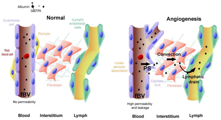
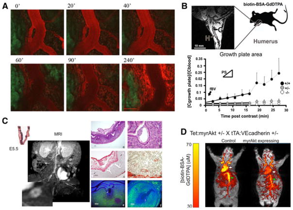
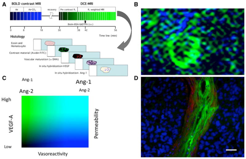
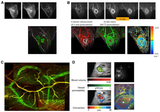
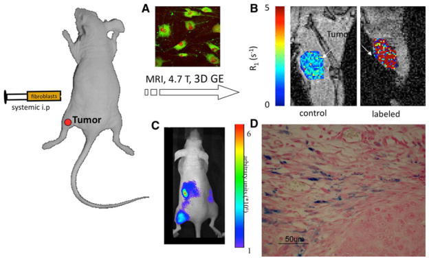
During the early stages of angiogenesis, following stimulation of endothelial cells by vascular endothelial growth factor (VEGF), the vascular wall is breached, allowing high molecular weight proteins to leak from the vessels to the interstitial space. This hallmark of angiogenesis results in deposition of a provisional matrix, elevation of the interstitial pressure and induction of interstitial convection. Albumin, the major plasma protein appears to be an innocent bystander that is significantly affected by these changes, and thus can be used as a biomarker for vascular permeability associated with angiogenesis. Traditionally, albumin leak in superficial organs was followed by colorimetry or morphometry with the use of albumin binding vital dyes. Over the last years, the introduction of tagged-albumin that can be detected by various imaging methods, such as magnetic resonance imaging and positron emission tomography, opened new possibilities for quantitative three dimension dynamic analysis of permeability in any organ. Using these tools it is now possible to follow not only vascular permeability, but also interstitial convection and lymphatic drain. Active uptake of tagged albumin by caveolae-mediated endocytosis opens the possibility for using labeled albumin for vital staining of cells and cell tracking. This approach was used for monitoring recruitment of perivascular stroma fibroblasts associated with tumor angiogenesis.


Figures

Fig. 1
Fig. 1
Measurement of vascular permeability, interstitial convection and lymphatic drain using tagged albumin. (Left) In normal tissue with mature blood vessels, after intravenous injection, albumin-GdDTPA does not extravasate, and its concentration inside the blood vessels slowly decreases (t1/2 = 4 h). (Right) During angiogenesis blood vessels are hyperpermeable. Initially, the contrast agent is only in the vessels, yielding an estimation of the microvascular density (fBV). The rate (or slope) of interstitial accumulation of the contrast agent provides the permeability surface area product (or permeability; PS). Elevated interstitial pressure, for example in the center of a tumor, can drive interstitial convection (convection). Eventually after a longer period (>60 min) the extravasated albumin-GdDTPA is drained from the interstitium into the lymphatics
Fig. 2
Fig. 2
MRI and fluorescence imaging of vascular permeability. a Confocal intravital microscopy of angiogenic vasculature of an MLS ovarian tumor implanted in a dorsal skin flap chamber. BSA-Rhodamine (ROX) was administered intravenously and followed for 4 h. Time points 0–90 min were acquired without moving the animal. Point 120 & 240 min were acquired after the animal was awake and re-anesthesized for continued imaging (bar, 100 micron). b Analysis of blood vessel permeability in the growing bone, showing difference between the permeable vessels in wild-type (+/+) mice relative to the non permeable vessels in PKBalpha/Akt1 heterozygous (±) or knockout (−/−) mice [51]. c Vascular permeability enhanced in sites of fetal implantation. (Left) MRI after administration of biotin-BSA-GdDTPA (inset; magnification of a single implantation site). (Right) histological analysis of implantation sites showing H&E staining (a, b), vascular alphaSMA staining of mature vessels (c, d) and histological analysis of the contrast media (e, f) [49]. d MRI analysis of systemic vascular expansion and elevated permeability induced by endothelial expression of activated PKBalpha/Akt1. Color scale, concentration of biotin-BSA-GdDTPA [69]
Fig. 3
Fig. 3
Multiparametric mapping of vascular permeability and vascular maturation. a Consecutive BOLD contrast response to hypercapnia and DCE-MRI followed by histological analysis can be used for mapping vascular maturation and permeability. b Map of vasoreactivity to hypercapnia (in blue) and vascular permeability to biotin-BSA-GdDTPA (in green) of an ovarian carcinoma xenograft, showing the high vasoreactivity and low permeability of the normal tissue and the high permeability and low vasoreactivity characteristic of the tumor vessels. c Vasoreactivity and vascular permeability as functional biomarkers for the activity of angiogenic growth factors such as the relative fraction of angiopoietins 1 and 2 that control vascular maturation and VEGF as a regulator of vascular permeability. d Fluorescence imaging of vascular permeability (green; avidin-FITC staining of biotin-BSA-GdDTPA) and maturation (red; alpha-SMA staining of pericytes, vascular smooth muscle cells and myofibroblasts) in histological section of an ovarian carcinoma tumor [39, 70]
Fig. 4
Fig. 4
Imaging permeability, convection and lymphatic drain using biotin-BSA-GdDTPA (FAM/ROX). a Delayed enhancement observed in a mouse in which biotin-BSA-GdDTPA was administered intravenously and allowed to circulate for 6 h (green) and 3 min (red). Contrast material extravasated from the VEGF over expressing tumor in the limb of the mouse and drained towards the popliteal lymph node. b In vivo avidin chase analysis of lymphatic clearance. Administration of avidin results in immediate clearance of the intravascular contrast media. Lower row: left, initial enhancement angiogram (red); center, avidin induced clearance (green); right, life time for interstitial albumin clearance [16, 30]. c Fluorescence imaging of lymphatic uptake of extravasated albumin (lymphatic vessels, biotin-BSA-GdDTPA-FAM, green; blood vessels, BSA-ROX, red). d Angiogenesis of ovarian grafts, showing high convection and lymphatic drain of extravasated biotin-BSA-GdDTPA [56, 57]
Fig. 5
Fig. 5
Recruitment of fibroblasts detected using tagged albumin. a Fluorescence microscopy of fibroblasts incubated with biotin-BSA-GdDTPA (green). b MRI analysis showing elevated R1 in subcutaneous ovarian carcinoma tumors when labeled fibroblasts were administered intraperitonealy and recruited to the tumor. c NIR imaging showing recruitment of DiR labeled fibroblasts. d Histological Prussian Blue staining showing recruitment of iron-oxide labeled fibroblasts to ovarian carcinoma tumors [61, 62]

Similar articles

Cited by

References

    1. Leung DW, Cachianes G, Kuang WJ, et al. Vascular endothelial growth factor is a secreted angiogenic mitogen. Science. 1989;246:1306–1309. - PubMed
    1. Dvorak HF. Discovery of vascular permeability factor (VPF) Exp Cell Res. 2006;312:522–526. - PubMed
    1. Dvorak HF, Nagy JA, Feng D, et al. Vascular permeability factor/vascular endothelial growth factor and the significance of microvascular hyperpermeability in angiogenesis. Curr Top Microbiol Immunol. 1999;237:97–132. - PubMed
    1. Dvorak AM, Feng D. The vesiculo-vacuolar organelle (VVO). A new endothelial cell permeability organelle. J Histochem Cytochem. 2001;49:419–432. - PubMed
    1. Nagy JA, Benjamin L, Zeng H, et al. Vascular permeability, vascular hyperpermeability and angiogenesis. Angiogenesis. 2008;11:109–119. - PMC - PubMed

Publication types