. 2010 Jul;341(1):95-110.
doi: 10.1007/s00441-010-0984-6. Epub 2010 May 30.

Neurod1 regulates survival and formation of connections in mouse ear and brain

Affiliations

Neurod1 regulates survival and formation of connections in mouse ear and brain

Israt Jahan et al. Cell Tissue Res. 2010 Jul.

Abstract

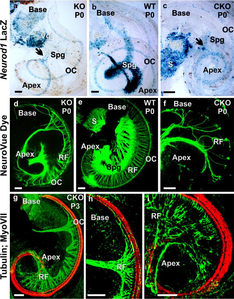
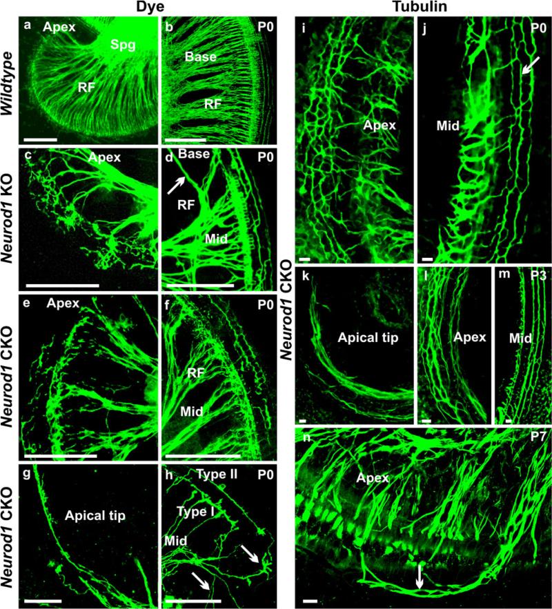
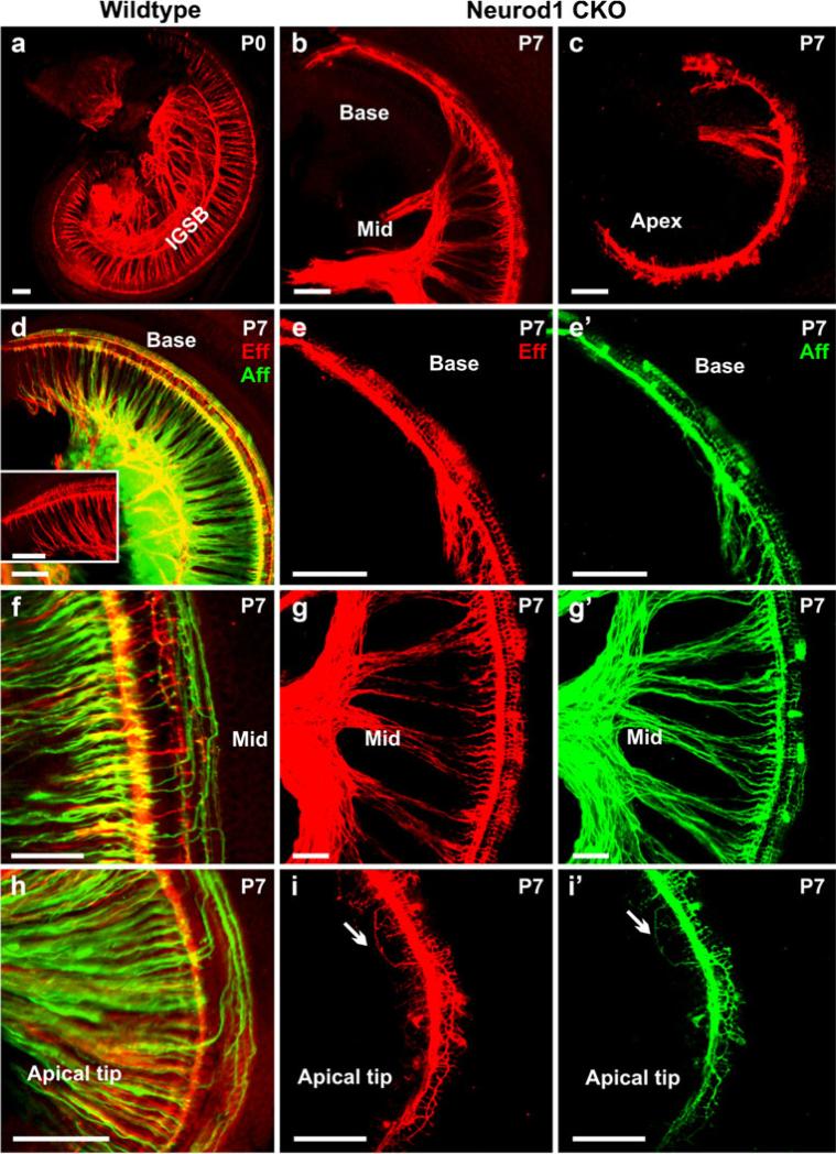
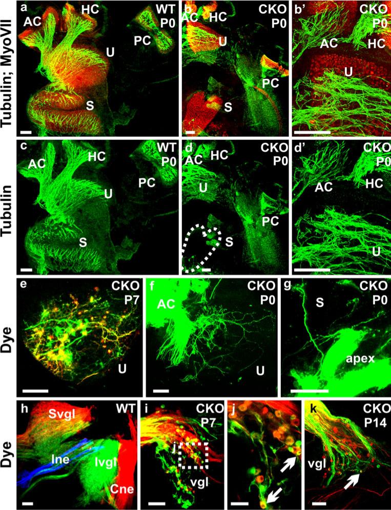
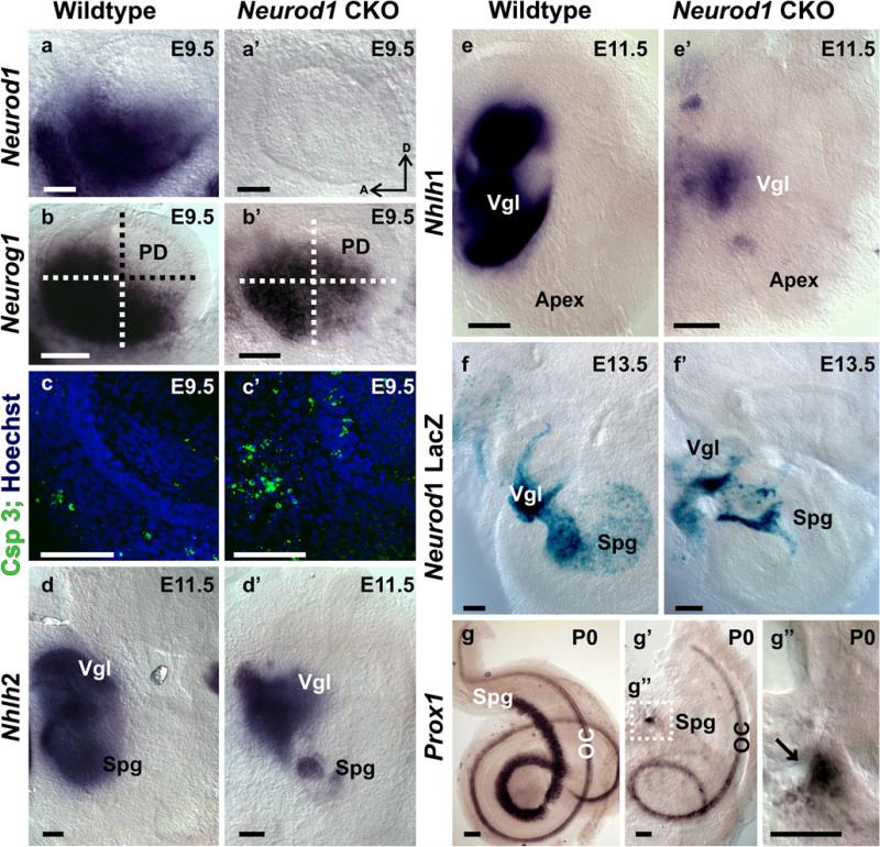
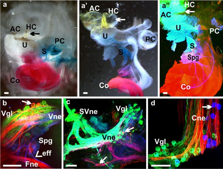
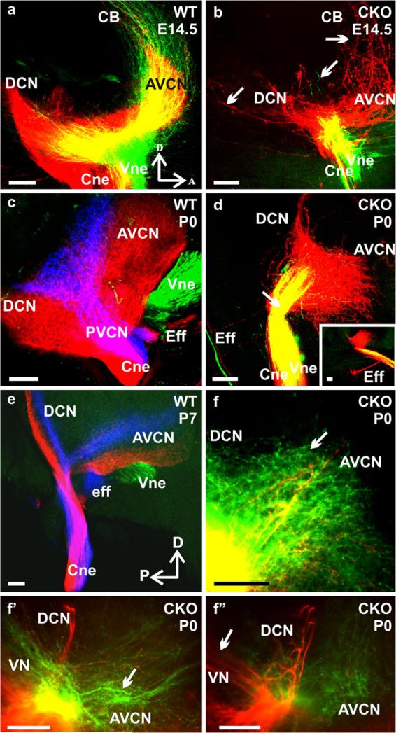
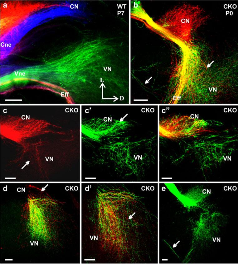
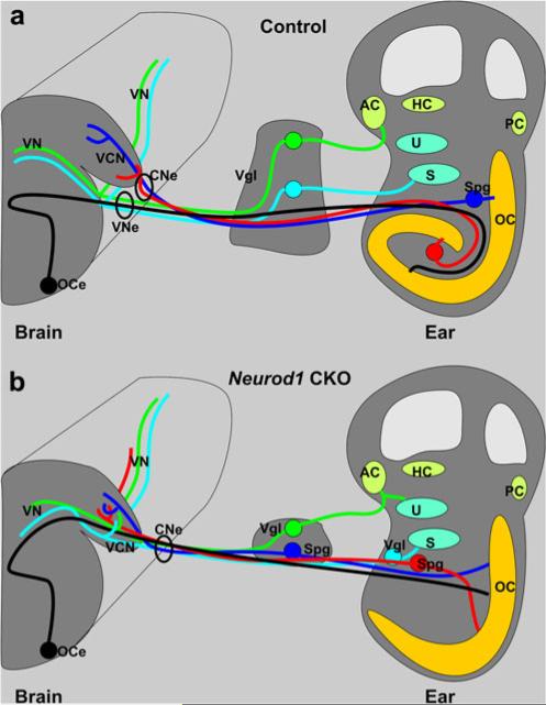
The developing sensory neurons of the mammalian ear require two sequentially activated bHLH genes, Neurog1 and Neurod1, for their development. Neurons never develop in Neurog1 null mice, and most neurons die in Neurod1 null mutants, a gene upregulated by Neurog1. The surviving neurons of Neurod1 null mice are incompletely characterized in postnatal mice because of the early lethality of mutants and the possible compromising effect of the absence of insulin on peripheral neuropathies. Using Tg(Pax2-cre), we have generated a conditional deletion of floxed Neurod1 for the ear; this mouse is viable and allows us to investigate ear innervation defects of Neurod1 absence only in the ear. We have compared the defects in embryos and show an ear phenotype in conditional Neurod1 null mice comparable with the systemic Neurod1 null mouse. By studying postnatal animals, we show that Neurod1 not only is necessary for the survival of most spiral and many vestibular neurons, but is also essential for a segregated central projection of vestibular and cochlear afferents. In the absence of Neurod1 in the ear, vestibular and cochlear afferents enter the cochlear nucleus as a single mixed nerve. Neurites coming from vestibular and cochlear sensory epithelia project centrally to both cochlear and vestibular nuclei, in addition to their designated target projections. The peripheral innervation of the remaining sensory neurons is disorganized and shows collaterals of single neurons projecting to multiple endorgans, displaying no tonotopic organization of the organ of Corti or the cochlear nucleus. Pending elucidation of the molecular details for these Neurod1 functions, these data demonstrate that Neurod1 is not only a major factor for the survival of neurons but is crucial for the development of normal ear connections, both in the ear and in the central nervous system.


Figures

Fig. 1
Fig. 1
Neurod1 conditional knockout (CKO) and knockout (KO) mice have a similar phenotype. In newborn mice, Neurod1-LacZ reaction shows expression in spiral ganglion (Spg) in wildtype (WT) mice (b), whereas only a few scattered spiral ganglion neurons are positive for Neurod1-LacZ in the Neurod1 KO and CKO (arrows in a, c). Additional expression of Neurod1-LacZ is seen in the hair cells of the organ of Corti (OC). The near complete loss of spiral ganglion neurons results in alterations of the projections of afferents in both mutants (d, f). The wildtype (e) has a well-developed spiral ganglion, and lipophilic dyes show regularly spaced radial fibers that carry both afferent and efferent fibers to the OC. In contrast, fewer and disorganized afferents reach the OC in both Neurod1 KO and CKO mice. The fiber reduction in the CKO mice is more severe in the apex and base, with some retained innervation in the middle turn, consistent with the region of remaining spiral ganglion neurons shown with tubulin (green) and Myo VII (red) immunofluorescence staining (g–i). (RF radial fibers, S saccule, Vgl vestibular ganglion). Bars 100 μm
Fig. 2
Fig. 2
Afferents are disorganized in Neurod1 mutant mice. Afferents form tightly spaced radial fiber bundles to the organ of Corti (a, b) in wildtype animals (RF radial fibers, Spg spiral ganglion). In Neurod1 KO and CKO mice, radial fibers are severely reduced and more widely spaced with profuse collateral branching (c–n). Both mutants have few recognizable type II fibers in the middle turn of the cochlea (d, m), but most of these fibers near the apical region are reduced or disorganized with random extension toward the base (arrow in d) or apex (f, i, arrow in j, l). The type I fibers have extensive branching to the inner hair cells near the lower middle turn (c, e, h, i, n). The basal and apical tips are innervated by a single fiber projecting from the inner spiral bundles with progressive abolition of type II fibers (g, k). Lipophilic dye applications to the apex of the Neurod1 CKO mice shows fibers in the middle turn bearing characteristics of type II fibers extending along the rows of outer hair cells (h). In addition, some fibers to the inner hair cells are labeled and resemble normal type I afferents, but many other fibers branch profusely (arrows in h) as they reach inner hair cells near the apical middle turn (h). This abnormal branching and overshooting of type I fibers in the apical region is observed more clearly in later stages in which they appear to innervate hair cells in the rows of outer hair cells (arrow in n). Bars 100 μm (a–h), 10 μm (i–n)
Fig. 3
Fig. 3
Defects in efferent innervation are comparable with those in afferents in Neurod1 CKO mice. Efferent fibers (red, Eff) in the Neurod1 CKO mice are massively reduced and disorganized, comparable with the afferent fibers (green, Aff) shown with dye application in the olivo-cochlear efferent bundles and cochlear nucleus. Only the wildtype mice show the formation of the intraganglionic spiral bundle (IGSB) together with the efferents to inner and outer hair cells (a, d, inset in d, f, h). The efferent organization is disrupted in the absence of Neurod1. Near the middle turn of Neurod1 CKO mice, some efferent fibers innervate the inner hair cells along with the afferent fibers (g, g’). A collateral branch from this efferent extends as a single fiber along type I afferent fiber to the inner hair cells without any formation of radial fibers (b, c, e, e’). In the apical tip, both efferents and afferents are reduced, with random overshooting of type II fibers, which extend toward the apex (arrows in i, i’). Some efferent fibers cross the tunnel of Corti innervating only the first row of outer hair cells rather than forming three parallel bundles along three rows of outer hair cells (b, c, e, g, i). The disorganization of afferents in the cochlea is even more severe in the later stage, with progressive reduction of type II neurons (e’, g’, i’). Bars 100 μm (a–e’, h–i’), 50 μm (f–g’)
Fig. 4
Fig. 4
Absence of Neurod1 causes reduction of innervation to vestibular end organs with more profound loss in the saccule (S). The innervation of vestibular end organs in Neurod1 CKO mice are reduced and disorganized (b, b’, d, d’, e) in comparison with the control littermate (a, c) shown with the merged tubulin (green) and Myo VII (red) immunofluorescence staining (HC horizontal crista, PC posterior crista). In addition, the abnormal branching of fibers to reach other sensory epithelia is observed by dye application to the anterior crista (AC); this results in fibers reaching the utricle (f, U). These fibers take unusual trajectories and form both calyceal and bouton endings on hair cells (f). Such branching is also obvious in dye applications in the apex of the cochlea which labels few fibers to the saccule (g). The vestibular ganglia (vgl) in Neurod1 CKO mice (i–k) are reduced in comparison with the control ganglia (h), which are labeled from the brainstem dye injection (Cne cochlear nerve, Ine intermediate nerve, Ivgl inferior vestibular ganglion, Svgl superior vestibular ganglion). Moreover, these ganglia represent mixed types of neurons, which label both the cochlear and vestibular nuclei (arrows in j, k). These mixed ganglia may therefore be responsible for fiber projection in between the different sensory epithelia (f, g). Bars 100 μm (a–i, k), 50 μm (j)
Fig. 5
Fig. 5
Conditional deletion of Neurod1 is early and complete and induces gene expression changes. In situ hybridization with a Neurod1 probe results in massive labeling in the otocyst as early as E9.5 in wildtype mice (a), whereas no Neurod1 in situ hybridization signal can be seen in Neurod1 CKO mice (a’). The bHLH gene Neurog1 shows changes in expression with upregulation in the postero-dorsal quadrant and a reduction in the ventral half (b, b’) in the absence of Neurod1 (PD postero-dorsal). These changes imply that Neurod1 has been effectively recombined at least a few hours before our investigation of these expression changes. The distribution of activated Caspase 3, a marker for apoptotic cell death, shows greater apoptotic cells both inside and even more profoundly outside the otocyst in the forming vestibular ganglion (c, c’). These data suggest that Neurod1 absence leads to a rapid loss of neuronal precursors through apoptosis and could be related to the signal reduction of Neurog1 in the ventral half (b’). Nhlh2 is another early marker of developing sensory neurons. This gene shows expression changes with a reduction in spiral ganglion (Spg) neurons as early as E11.5 (d, d’; Vgl vestibular ganglion). Similar changes are also seen in another early neuronal marker, Nhlh1, which makes the loss of sensory neurons expression more obvious (e, e’). A direct comparison of wildtype and Neurod1 CKO or KO (shown here) mice shows that, by E13.5, the loss of spiral ganglion neurons is completed (f, f’) validating the observations of an early onset of apoptosis. A specific marker for spiral ganglion neurons, Prox1, confirms the loss of spiral ganglia in a later stage (g’, g”; OC organ of Corti). Bars 100 μm
Fig. 6
Fig. 6
Afferent disorganization may correlate with the mixed ganglia in Neurod1 CKO mice. Afferents of the inner ear were labeled by using filter strips that were soaked with chromatically different lipophilic dye tracers and that were inserted into the various sensory epithelia of inner ear (AC anterior crista, HC horizontal crista, PC posterior crista, U utricle, S saccule, Co cochlea, Spg spiral ganglion, Vgl vestibular ganglion, Cne cochlear nerve, Vne vestibular nerve, Fne facial nerve, Eff efferent fibers, SVne superior vestibular nerve). NeuroVue red (direct and false-colored red) dye was applied into the apex of cochlea, with NeuroVue jade (direct color yellow, false-colored green; arrows in a–a”) dye into the anterior canal crista/horizontal canal crista/utricle and NeuroVue maroon (direct and false-colored blue) into the base of cochlea or into the saccule (a-a”). Vestibular ganglion neurons with vestibular nerve fibers were labeled by the dye inserted into the canal crista and were shown to branch unusually from the vestibular nerve to innervate the canal crista and utricle (c). In addition, these ganglion neurons were labeled not only from the vestibular end organs, but also from the dye injected into the cochlea (red neurons, arrow in b, c). These mixed ganglion neurons migrated unusually near the nerve entry site to the brainstem (arrow in d). Moreover, efferents entered into the ear together with the facial nerve (arrowhead in b). Bars 100 μm
Fig. 7
Fig. 7
Loss of Neurod1 results in overlapping central projections. Various dyes were applied into the different sensory epithelia of the inner ear to label their central projections. This approach showed segregated central projections of vestibular and cochlear afferents to the vestibular and cochlear nuclei and other targets such as the cerebellum as early as E14.5 (a). In contrast, afferent fibers entered into the brain as a single root in Neurod1 CKO mice (b, d, f-f”), and afferents labeled from the cochlea projected to the cerebellum instead of stopping at the antero-ventral cochlear nucleus (b). In newborn and P7 wildtype mice, central projections from the base (blue), apex (red), and vestibular endorgans (green) and all afferents entered the hindbrain as discrete fascicles, terminating exclusively in cochlear and vestibular nuclei (c, e). In Neurod1 CKO mice, afferents to the cochlear nuclei were greatly reduced, showing overlapping projections from both the apex and base, with little extension toward the dorsal cochlear nucleus, and both vestibular and cochlear afferents entered through the same root (arrow in d). Vestibular afferents not only entered through the cochlear nucleus, but also projected variably to the antero-ventral cochlear nucleus (arrow in f) in addition to the vestibular nucleus (f’). Similarly cochlear afferents projected to vestibular nucleus (arrow in f”) in addition to cochlear nucleus.. Efferent fibers to the ear normally exited with the vestibular nerve root (c) but instead exited through the facial nerve in the mutant (d, insert in d) and were rerouted outside the brain to the vestibulo-cochlear nerve or the “cochlear nerve root” (AVCN antero-ventral cochlear nucleus, PVCN postero-ventral cochlear nucleus, DCN dorsal cochlear nucleus, CB cerebellum, Cne cochlear nerve, Vne vestibular nerve, Eff efferent nerve fibers, VN vestibular nucleus, A anterior, D dorsal, P posterior). Bars 100 μm
Fig. 8
Fig. 8
Vestibular and cochlear fibers project to both nuclei in Neurod1 CKO mice. Coronal sections from the cochlear and vestibular nuclei with same dye injections as shown in Fig. 7 reveal that afferents labeled from the cochlea (red) target mostly the cochlear nucleus but extend also to the vestibular nuclei (right arrow in b, arrows in c, d, d’) in the absence of Neurod1. Likewise, afferents labeled from vestibular endorgans (green) project into the cochlear nucleus in addition to their original vestibular nuclei projections (b, c’, c”, d, d’, e). This altered and overlapping projection is confirmed by specific dye application only in vestibular or cochlear epithelia showing that the fibers from vestibular end organs reach to the cochlear nucleus and vice versa (vestibular afferents labeling shown in e). The sections from wildtypes demonstrate the distinct topology of afferents from vestibular epithelia (green), cochlear apex (red), and cochlear base (blue) and the efferents that exit with the vestibular nerve root (a), whereas they exit through the cochlear nerve root (b) or through the facial nerve (left arrows in b, arrow in e) in Neurod1 CKO mice (CN cochlear nucleus, Cne cochlear nerve, Eff efferent nerve fibers, VN vestibular nucleus, Vne vestibular nerve, L lateral, D dorsal). Bars 100 μm
Fig. 9
Fig. 9
Illustration comparing the neuronal connection of the wildtype ear with the brain (a) with that of the Neurod1 conditional null (CKO) mouse (b). All major features can be distinguished, but the cochlea is shortened and misshapen in Neurod1 CKO mice. Whereas the normal organ of Corti (OC) has multiple turns and an accompanying spiral ganglion (Spg), there is typically no spiral ganglion found near the shortened organ of Corti in Neurod1 CKO mice. However, spiral ganglion neurons as verified by their backfilling from the organ of Corti can be found near the saccule (S) or in the much reduced vestibular ganglion. A comparable but less severe disorganization is found in the vestibular ganglia (Vgl), which normally lie outside the ear but are scattered both inside and outside the ear in Neurod1 CKO mice. As a consequence, nearby neurons may project to both vestibular and cochlear sensory epithelia. As fibers approach the brain, spiral afferents normally segregate from vestibular afferents, so that each fascicle enters as distinct vestibular and cochlear nerves into the vestibular and cochlear nuclei (a). In contrast, in Neurod1 CKO mice, all afferents enter the brain together as a single root into the ventral cochlear nucleus (VCN). Despite this common entry point, fibers nevertheless project to the vestibular nuclei (VN) but also end up without any apparent topology in the cochlear nuclei. These data show that Neurod1 expression is needed for proper migration of peripheral and central projection of all spiral neurons and to a lesser extent of vestibular neurons. Rerouting to the periphery is also observed in olivocochlear and vestibular efferents, which normally exit the brain via the vestibular nerve to reroute to the cochlea but instead exit the brain via the cochlear nerve or, less often, via the facial nerve to enter into the ear (AC anterior crista, HC horizontal crista, PC posterior crista, U utricle, CNe cochlear nerve, OCe olivocochlear efferent, VNe vestibular nerve)

Similar articles

Cited by

References

    1. Bermingham-McDonogh O, Oesterle EC, Stone JS, Hume CR, Huynh HM, Hayashi T. Expression of Prox1 during mouse cochlear development. J Comp Neurol. 2006;496:172–186. - PMC - PubMed
    1. Bermingham NA, Hassan BA, Price SD, Vollrath MA, Ben-Arie N, Eatock RA, Bellen HJ, Lysakowski A, Zoghbi HY. Math1: an essential gene for the generation of inner ear hair cells. Science. 1999;284:1837–1841. - PubMed
    1. Chen P, Johnson JE, Zoghbi HY, Segil N. The role of Math1 in inner ear development: uncoupling the establishment of the sensory primordium from hair cell fate determination. Development. 2002;129:2495–2505. - PubMed
    1. Cowan CA, Yokoyama N, Bianchi LM, Henkemeyer M, Fritzsch B. EphB2 guides axons at the midline and is necessary for normal vestibular function. Neuron. 2000;26:417–430. - PubMed
    1. Dabdoub A, Puligilla C, Jones JM, Fritzsch B, Cheah KS, Pevny LH, Kelley MW. Sox2 signaling in prosensory domain specification and subsequent hair cell differentiation in the developing cochlea. Proc Natl Acad Sci USA. 2008;105:18396–18401. - PMC - PubMed

Publication types

MeSH terms

Substances

LinkOut - more resources