. 2010 Jul;120(1):55-66.
doi: 10.1007/s00401-010-0702-1. Epub 2010 May 30.

Phosphorylated TDP-43 pathology and hippocampal sclerosis in progressive supranuclear palsy

Affiliations

Phosphorylated TDP-43 pathology and hippocampal sclerosis in progressive supranuclear palsy

Osamu Yokota et al. Acta Neuropathol. 2010 Jul.

Abstract

TDP-43 is characteristically accumulated in TDP-43 proteinopathies such as frontotemporal lobar degeneration and motor neurone disease, but is also present in some tauopathies, including Alzheimer's disease, argyrophilic grain disease, and corticobasal degeneration (CBD). However, several studies have suggested that cases of progressive supranuclear palsy (PSP) lack TDP-43 pathology. We have therefore examined limbic regions of the brain in 19 PSP cases, as well as in 12 CBD cases, using phosphorylation-dependent anti-TDP-43 antibodies. We observed TDP-43-positive inclusions in five PSP cases (26%), as well as in two CBD cases (17%). The amygdala and hippocampal dentate gyrus were most frequently affected in PSP. Regional tau burden tended to be higher in TDP-43-positive PSP cases, and a significant correlation between tau and TDP-43 burden was noted in the occipitotemporal gyrus. Hippocampal sclerosis (HS) was found in 3/5 TDP-43-positive PSP cases, but HS was significantly more frequent in TDP-43-positive than TDP-43 negative PSP cases. Dementia was present in 13/19 (58%) of the PSP cases, in 4/5 TDP-43-positive cases, in all 3 TDP-43-positive cases with HS, in 1/2 TDP-43-positive cases without HS, and 7/14 cases lacking both. TDP-43 and tau were frequently colocalized in the amygdala, but not in the hippocampal dentate gyrus. Immunoblotting demonstrated the characteristic (for TDP-43 proteinopathies) 45 and 25 kDa bands and high molecular weight smear in the TDP-43-positive PSP case. These findings suggest that (1) although PSP is nominally a tauopathy, pathological TDP-43 can accumulate in the limbic system in some cases, and (2) TDP-43 pathology may be concurrent with HS.


Figures
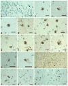
Fig. 1
Fig. 1
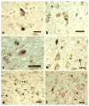
TDP-43-positive lesions in PSP. a Neuronal cytoplasmic inclusions (NCIs) in the hippocampal dentate gyrus. bd NCIs in the entorhinal cortex. Irregular shaped NCIs in the entorhinal cortex (e), fusiform gyrus (f), and subiculum (g). These inclusions have weakly stained or unstained regions. Small dot-like structures are also seen in the neuronal cytoplasm (g). Horseshoe-shaped (h, i) and NFT-like (j) NCIs in the entorhinal cortex. Intranuclear inclusions in the amygdala (k) and in the subiculum (l), cases PSP3 and PSP2, respectively. m Massive short threads-like structures in the subiculum, case PSP3. n Thick, thread-like structures in the amygdala. o, p Long, thin thread-like structures in the amygdala. pAb pS409/410 immunohistochemistry. All scale bars 20 μm
Fig. 2
Fig. 2
TDP-43-positive lesions in CBD. a Neuronal cytoplasmic inclusions (NCIs) in CA3 region of hippocampus. b, c Neuronal intranuclear inclusions in the amygdala. d A thick neurite and thin, thread-like structures in the amygdala. e Short thread-like structures and glial cytoplasmic inclusions (GCIs) in the alveus in the entorhinal cortex. f Coiled body-like structures and GCis in the alveus in the entorhinal cortex. pAb pS409/410 immunohistochemistry. All scale bars 20 μm
Fig. 3
Fig. 3
Tau burden in the limbic system in PSP cases with and without TDP-43 pathology. In all regions but the subiculum, tau burden in PSP cases with TDP-43 pathology is more severe than that in PSP cases without TDP-43 pathology. Stage 0–1, no to mild tau deposition; stages 2–3, moderate to severe tau deposition; stage 4, very severe tau deposition (see detailed definition in the text). TDP-43+ TDP-43-positive, TDP-43− TDP-43-negative, Amy amygdala, antERC the anterior portion of the entorhinal cortex, DG hippocampal dentate gyrus, Subi subiculum, postERC the posterior portion of the entorhinal cortex, FG fusiform gyrus, OTG occipitotemporal gyrus
Fig. 4
Fig. 4
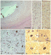
Pathological features in the hippocampus in a PSP case with TDP-43, HS, and argyrophilic grains (PSP5). a A low power view of the hippocampal CA1 to subiculum. Severe reduction of the width with tissue rarefaction is noted in the subiculum (arrow) and to a lesser degree in the adjacent CA1 region (arrowhead). b A moderate power view of the subiculum on the same section as that shown in a. Severe neuronal loss associated with gliosis is evident. Argyrophilic threads and grains are scattered, but tangles are rare. c The subiculum on an adjacent section of b. A moderate number of tau-positive threads and grains, but only a few tangles, are seen. d Argyrophilic grains in CA1 region. e, f TDP-43-positive cytoplasmic inclusions in CA1 region. g An irregular shaped TDP-43 accumulation in the subiculum. h A coiled body-like TDP-43-positive inclusion in the subiculum. a, b, d Gallyas-Braak hematoxylin-eosin stain. c AT-8 immunohistochemistry. eh pAb pS409/410 immunohistochemistry. Scale bars a 400 μm, b, c 25 μm, d 50 μm, eh 20 μm
Fig. 5
Fig. 5
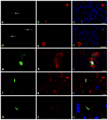
Confocal double-imunofluorescence of TDP-43 (a, d, g, j, m) and tau (b, e, h, k, n) in PSP cases. Merged images are shown in c, f, i, l, and o. Blue fluorescence in merged images are nuclei. af In the hippocampal dentate gyrus, TDP-43 accumulation (arrows) is not colocalized with tau labeling. gi In the amygdala, TDP-43 accumulation is often intermingled and colocalized with neuronal tau accumulation. jo TDP-43-positive neurites (j, m) and many tau-positive neurites and granules (k, n) are seen in the amygdala. Coexistence of TDP-43 and tau is noted in some neurites (l, o). AT8 and pAb pS409/410 double immunofluorescence. Scale bars ac 25 μm, df 25 μm, gi 2.5 μm, jl 7.5 μm, mo 7.5 μm
Fig. 6
Fig. 6
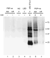
Immunoblot analysis of the sarkosyl-insoluble fraction in representative PSP cases with phosphorylation-dependent monoclonal anti-TDP-43 antibody (mAb pS409/410). The 45 kDa full length TDP-43, 25 kDa fragments, and high molecular weight smear are strongly labeled in the amygdala of a PSP case with TDP-43 pathology (lane 6) and in the frontal cortex of a FTLD-TDP case (lane 5). Weakly stained 45 and 25 kDa bands are noted in the hippocampus of a PSP case (lane 7), in which TDP-43 pathology was mild. Similar 45 and 25 kDa bands and smears were not immunolabeled in any of the other cases without detectable TDP-43 pathology by immunohistochemistry (lanes 1–4). Normal 43 kDa TDP-43 is not stained by this phophorylation-dependent antibody in any case. PSP progressive supranuclear palsy, LBD Lewy body disease, NC normal control, AM amygdala, HP hippocampus, F frontal cortex, T temporal cortex, IHC pAb pS409/410 immunohistochemistry

Similar articles

Cited by

References

    1. Ala TA, Beh GO, Frey WH., 2nd Pure hippocampal sclerosis: a rare cause of dementia mimicking Alzheimer’s disease. Neurology. 2000;54:843–848. - PubMed
    1. Amador-Ortiz C, Lin WL, Ahmed Z, et al. TDP-43 immunoreactivity in hippocampal sclerosis and Alzheimer’s disease. Ann Neurol. 2007;61:435–445. - PMC - PubMed
    1. Arai T, Hasegawa M, Akiyama H, et al. TDP-43 is a component of ubiquitin-positive tau-negative inclusions in frontotemporal lobar degeneration and amyotrophic lateral sclerosis. Biochem Biophys Res Commun. 2006;351:602–611. - PubMed
    1. Arai T, Mackenzie IR, Hasegawa M, et al. Phosphorylated TDP-43 in Alzheimer’s disease and dementia with Lewy bodies. Acta Neuropathol. 2009;117:125–136. - PubMed
    1. Beach TG, Sue L, Scott S, et al. Hippocampal sclerosis dementia with tauopathy. Brain Pathol. 2003;13:263–278. - PMC - PubMed

Publication types

MeSH terms