Review
. 2010 Jun 1;110(3):545-53.
doi: 10.1002/jcb.22574.

Beta-catenin--a supporting role in the skeleton

Affiliations
Review

Beta-catenin--a supporting role in the skeleton

Natasha Case et al. J Cell Biochem. .

Abstract

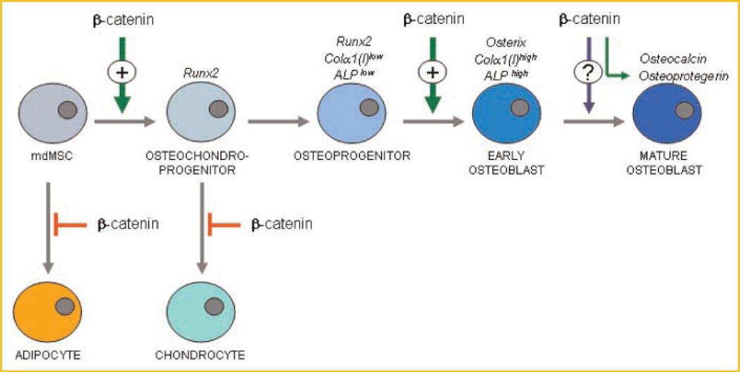
In the last 5 years a role for beta-catenin in the skeleton has been cemented. Beginning with mutations in the Lrp5 receptor that control beta-catenin canonical downstream signals, and progressing to transgenic models with bone-specific alteration of beta-catenin, research has shown that beta-catenin is required for normal bone development. A cell critical to bone in which beta-catenin activity determines function is the marrow-derived mesenchymal stem cell (MSC), where sustained beta-catenin prevents its distribution into adipogenic lineage. beta-Catenin actions are less well understood in mature osteoblasts: while beta-catenin contributes to control of osteoclastic bone resorption via alteration of the osteoprotegerin/RANKL ratio, a specific regulatory role during osteoblast bone synthesis has not yet been determined. The proven ability of mechanical factors to prevent beta-catenin degradation and induce nuclear translocation through Lrp-independent mechanisms suggests processes by which exercise might modulate bone mass via control of lineage allocation, in particular, by preventing precursor distribution into the adipocyte pool. Effects resulting from mechanical activation of beta-catenin in mature osteoblasts and osteocytes likely modulate bone resorption, but whether beta-catenin is involved in osteoblast synthetic function remains to be proven for both mechanical and soluble mediators. As beta-catenin appears to support the downstream effects of multiple osteogenic factors, studies clarifying when and where beta-catenin effects occur will be relevant for translational approaches aimed at preventing bone loss and terminal adipogenic conversion.


Figures

Fig. 1
Fig. 1
Mechanical strain activates β-catenin translocation. Osteoblast cells were subjected to strain for 15–60 min and immunostained for active β-catenin (top) and nuclei stained with DAPI (middle). Images of active β-catenin and DAPI staining were merged (bottom) to show active β-catenin had translocated to the nucleus. Increased nuclear active β-catenin was detected 15 min after beginning mechanical strain by confocal microscopy. Adapted from Case et al. [2008].
Fig. 2
Fig. 2
β-catenin activation by mechanical factors is both direct and indirect. β-catenin signaling is repressed both by sequestration with cadherins and a-catenin at the membrane and by interactions with a multiprotein destruction complex, which targets cytosolic catenin for proteosomal degradation upon phosphorylation by GSK3β. Mechanical signals (strain/deformation, fluid shear, cell acceleration/vibration) in bone cells inhibit GSK3β activity, with Akt being a likely effector by phosphorylation of GSK3β at serine-9. Rapid Akt phosphorylation after mechanical input may occur directly via ILK with involvement of FAK, or more gradually through receptor-dependent actions on PI3K. Prolonged mechanical signaling may also enhance binding of Wnt to its co-receptors, leading to disruption of the destruction complex. Once released from alternative fates such as sequestration, complex binding, or degradation, β-catenin will translocate to the nucleus, binding to Tcf/Lef transcription factors and triggering cell processes.
Fig. 3
Fig. 3
β-catenin is activated by strain and is necessary to transmit both mechanical and pharmacological signals that result in inhibition of adipogenesis. A: MSCs in adipogenic medium were analyzed after exposure to daily strain regimen for 4 days, showing that both active and total β-catenin are increased by strain. During this time adiponectin, representing adipocyte conversion, is limited by mechanical strain. Note also that COX2 was induced by strain. B: MSCs were treated with siRNA scrambled (siScr) or targeting β-catenin (siCat) prior to beginning the strain regimen. Strain was unable to prevent adipogenesis when β-catenin was knocked down (densitometry shown below). β-catenin was not necessary for strain induction of COX2. C: Similarly, when adipogenesis was prevented by inhibiting GSK3β with the small molecule SB415286 (20 mm), siRNA targeting β-catenin interfered with this effect. The pharmacologic effect to induce COX2, again, did not require β-catenin to be effective. Adapted from Sen et al. [2009].
Fig. 4
Fig. 4
β-catenin restricts MSC distribution to non-osteoprogenitor lineages. Effects of β-catenin availability and activation prevent loss of precursor cells to the adipogenic phenotype. Once the MSC has become an osteochondroprogenitor, decreases in β-catenin allow diversion to the chondrocytic lineage. While elevated β-catenin appears to support the mature osteogenic phenotype, it has yet not been shown to be a singular impetus for this differentiation pathway.

Similar articles

Cited by

References

    1. Akune T, Ohba S, Kamekura S, Yamaguchi M, Chung UI, Kubota N, Terauchi Y, Harada Y, Azuma Y, Nakamura K, Kadowaki T, Kawaguchi H. PPARgamma insufficiency enhances osteogenesis through osteoblast formation from bone marrow progenitors. J Clin Invest. 2004;113:846–855. - PMC - PubMed
    1. Armstrong VJ, Muzylak M, Sunters A, Zaman G, Saxon LK, Price JS, Lanyon LE. Wnt/beta-catenin signaling is a component of osteoblastic bone cells’ early responses to load-bearing, and requires estrogen receptor alpha. J Biol Chem. 2007;282:20715–20727. - PubMed
    1. Bodine PV, Zhao W, Kharode YP, Bex FJ, Lambert AJ, Goad MB, Gaur T, Stein GS, Lian JB, Komm BS. The Wnt antagonist secreted frizzled-related protein-1 is a negative regulator of trabecular bone formation in adult mice. Mol Endocrinol. 2004;18:1222–1237. - PubMed
    1. Boyden LM, Mao J, Belsky J, Mitzner L, Farhi A, Mitnick MA, Wu D, Insogna K, Lifton RP. High bone density due to a mutation in LDL-receptor-related protein 5. N Engl J Med. 2002;346:1513–1521. - PubMed
    1. Brunkow ME, Gardner JC, Van Ness J, Paeper BW, Kovacevich BR, Proll S, Skonier JE, Zhao L, Sabo PJ, Fu Y, Alisch RS, Gillett L, Colbert T, Tacconi P, Galas D, Hamersma H, Beighton P, Mulligan J. Bone dysplasia sclerosteosis results from loss of the SOST gene product, a novel cystine knot-containing protein. Am J Hum Genet. 2001;68:577–589. - PMC - PubMed

Publication types