. 2010 Jun 1;110(3):671-86.
doi: 10.1002/jcb.22578.

Analysis of hairless corepressor mutants to characterize molecular cooperation with the vitamin D receptor in promoting the mammalian hair cycle

Affiliations

Analysis of hairless corepressor mutants to characterize molecular cooperation with the vitamin D receptor in promoting the mammalian hair cycle

Jui-Cheng Hsieh et al. J Cell Biochem. .

Abstract

The mammalian hair cycle requires both the vitamin D receptor (VDR) and the hairless (Hr) corepressor, each of which is expressed in the hair follicle. Hr interacts directly with VDR to repress VDR-targeted transcription. Herein, we further map the VDR-interaction domain to regions in the C-terminal half of Hr that contain two LXXLL-like pairs of motifs known to mediate contact of Hr with the RAR-related orphan receptor alpha and with the thyroid hormone receptor, respectively. Site-directed mutagenesis indicates that all four hydrophobic motifs are required for VDR transrepression by Hr. Point mutation of rat Hr at conserved residues corresponding to natural mutants causing alopecia in mice (G985W and a C-terminal deletion DeltaAK) and in humans (P95S, C422Y, E611G, R640Q, C642G, N988S, D1030N, A1040T, V1074M, and V1154D), as well as alteration of residues in the C-terminal Jumonji C domain implicated in histone demethylation activity (C1025G/E1027G and H1143G) revealed that all Hr mutants retained VDR association, and that transrepressor activity was selectively abrogated in C642G, G985W, N988S, D1030N, V1074M, H1143G, and V1154D. Four of these latter Hr mutants (C642G, N988S, D1030N, and V1154D) were found to associate normally with histone deacetylase-3. Finally, we identified three regions of human VDR necessary for association with Hr, namely residues 109-111, 134-201, and 202-303. It is concluded that Hr and VDR interact via multiple protein-protein interfaces, with Hr recruiting histone deacetylases and possibly itself catalyzing histone demethylation to effect chromatin remodeling and repress the transcription of VDR target genes that control the hair cycle.


Figures

Fig. 1
Fig. 1. C-terminal fragments of Hr associate with immobilized VDR
GST pull-downs were carried out as described in Methods. The following radiolabeled rHr fragments were generated by IVTT: an N-terminal portion with residues 31–568, a C-terminal portion containing 568–1207, and C-terminal fragments 450–730, 750–1084, 750–864, 864–981 and 980–1084. A: Inputs shown are 6.9% of the amounts used in the pull-down reactions. B: Pull-downs in which the IVTT-synthesized Hr fragments described above were incubated with immobilized GST-VDR, containing wild-type VDR. C: As a negative control, the radiolabeled Hr fragments were also incubated with immobilized GST alone. To allow for direct comparisons, the gels shown in panels A–C were exposed to film for the same amount of time. D: Tabular summary of pull-down (PDA) results in A–C for each rHr fragment appears to the right of a schematic diagram of rHr and its fragments. The ability of each Hr fragment to interact with VDR by coimmunoprecipitation (CoIP) [Hsieh et al., 2003] or by repressing (Fold Repress.) VDR signaling [Hsieh et al., 2003] also is listed to the right of the schematic. The location of the following Hr functional domains is depicted in the schematic: three repression domains (RD1-3) [Potter et al., 2001], interaction domains 1 and 2 (ID1, ID2) mediating interaction between Hr and the RAR-related orphan receptor-alpha (RORα) [Moraitis et al., 2002] or with the thyroid hormone receptor (TR) [Potter et al., 2002], and a Jumonji C-like domain (abbreviated JmC) [Clissold and Ponting, 2001].
Fig. 2
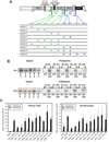
Fig. 2. Functional analysis of rat Hr point mutations in the interaction domains for RORα and TR
A: Various combinations of ID point mutations were used to create nine rHr mutants for functional testing, designated mutant 1 through mutant 9. Altered amino acids (all to alanine) are indicated by asterisks (*) and color-coded to match their corresponding interaction domains. Functional domains, in addition to those shown in Fig. 1D, are a nuclear localization domain [Djabali et al., 2001] and a zinc-finger domain [Cachon-Gonzalez et al., 1994]. B: GST pull-down results for the association of nuclear receptor binding domain mutants of Hr with immobilized VDR. The left panels of B show 4.5% of the inputs of 35S-labeled wild type (wt) or mutant (m1-m9) Hr proteins produced in an in vitro transcription-translation system (see Methods). The right panels of B illustrate pull-down experiments using labeled Hr proteins incubated with either immobilized GST protein (negative control) or GST-VDR fusion protein containing full-length human VDR. C: Ability of each mutant to repress VDR signaling. Expression plasmids encoding all nine mutants were transfected into COS-7 monkey kidney cells (left panel in C) or into human KERTr-1106 skin cells (right panel in C) as described in Methods. Mock transfected wells received reporter plasmid but no expression plasmid for VDR. Cells were treated with ethanol or 10 nM 1,25(OH)2D3 and allowed to incubate for 24 hours (COS-7) or 36 hours (KERTr-1106). Transcriptional activities were quantified by a luciferase assay and are normalized to the expression of Renilla luciferase, a constitutively expressed reporter gene used to monitor transcription efficiency. Error bars represent standard deviations for triplicate analyses. To obtain relative values for plotting of the KERTr-1106 results (right panel of C), transactivation units (ratios) obtained with wild type VDR and pRK5 (empty vector) in the presence of 1,25(OH)2D3 and the absence of Hr were arbitrarily set to 1.0 in six independent experiments (thus lacking an error bar); results for all Hr mutants are expressed in comparison to this arbitrary standard. The values are shown as an average of six determinations ± the standard deviation.
Fig. 3
Fig. 3. Testing of two alopecic mouse mutations in the context of rat Hr
A: The location of the two mouse alopecic mutations on either side of the conserved Jumonji C domain. The two mutations, originally described in mice [Brancaz et al., 2004; Nam et al., 2006], were recreated in rHr as G985W and Δ1206-1207 (designated ΔAK to denote the loss of the two terminal amino acids Ala-Lys). The conservation of the residues affected by these mutations, along with immediately flanking residues, is shown in the lower portion of panel A. Species abbreviations are: human (hum), mouse Mus musculus (mus), horse, Equus caballus (hrs), and opossum Monodelphis domestica (ops). Accession numbers for these sequences are as follows: rHr NP_077340.2; mouse Hr NP_068677.2; hHr NP_005135.2; dog (Canis lupus familiaris) Hr XP_543256.2 (predicted); pig (Sus scrofa) Hr NP_001077399.1; cow (Bos taurus) Hr NP_001096005.1; horse (Equus caballus) Hr XP_001490941.1; opossum Hr XP_001381979.1 (predicted); rat JmJD1a, also known as JmjC-containing histone demethylase 2A (JHDM2A) or as testis-specific gene A (TSGA), NP_786940; mouse JmJD1a, NP_766589; human JmJD1a, NP_060903.2; rat JmJD1b, also known as nuclear protein 5qNCA or as JHDM2B, XP_001061636 (predicted); mouse JmJD1b, NP_001074725; Human JmJD1b, NP_057688; rat JmJD1c, also called thyroid receptor interacting protein 8 (TRIP8) or JHDM2C, XP_001080424 (predicted); Mouse JmJD1c XP_980927 (predicted); and human JmJD1c NP_004232. B: GST-VDR pull-down analysis of the G985W and ΔAK rHr mutants using radiolabeled rHr proteins generated in an in vitro transcription-translation system as described in Methods. Beads contained either GST alone or full-length hVDR fused to GST (GST-VDR). Input lanes 1–4 correspond to, and represent 5% of the amount used for respective pull-down lanes 5/6, 7/8, 9/10, and 11/12.
Fig. 4
Fig. 4. Ability of the G985W and ΔAK rat Hr mutants to repress VDR- and TR-mediated transactivation of reporter genes
Expression plasmids for wild-type and mutant rHrs were cotransfected into COS-7 cells along with, if indicated, a reporter plasmid for VDR (the p24OHaseLuc vector) and the pSG5-hVDR expression plasmid (panels A and C) or a reporter plasmid for TR, (rMHC)2pLucMCS, and the pSG5-hTRB1 expression plasmid for human TRβ1 (B, D). Transfected cultures were treated ± 10 nM 1,25(OH)2D3 or ± 10 nM tri-iodothyronine (T3) as indicated. Luciferase assays were performed as described in Methods and the results shown represent the average of six replicates ± the standard deviation.
Fig. 5
Fig. 5. Functional characterization of selected rat Hr mutants which recapitulate naturally occurring point mutations in human Hr that confer alopecia
A: Loss of VDR corepressor activity by rHr mutants D1030N and V1154D. COS-7 cells were transfected with the natural VDRE-reporter plasmid p24OHaseLuc, pSG5-hVDR and an expression plasmid for the indicated rHr mutant as described in Methods. Cells were treated for 24 hrs ± 10 nM 1,25(OH)2D3 as indicated, and transcriptional activity was quantified via luciferase assay as described in Methods and the legend to Fig. 2C. Error bars represent standard deviations for triplicate wells. B: Radiolabeled rHr mutants were produced in an in vitro transcription-translation system, incubated with FLAG-tagged HDAC3 [Wen et al., 2000], immunoprecipitated with the indicated antisera, and subjected to SDS-PAGE and autoradiography as described in Methods. A small aliquot of radiolabeled input was electrophoresed in lanes indicated by “In”. The appearance of a labeled rHr band in the α-Flag lane, but not the non-specific IgG lane, is taken as evidence of a direct interaction between the rHr mutant and HDAC3.
Fig. 6
Fig. 6. Mutation of conserved residues in the Jumonji C domain of rat Hr and the functional consequence of these alterations on the VDR corepressor activity of Hr
A: Conservation of the three mutated residues which are postulated to confer enzymatic activity, such as histone demethylase, onto the Hr protein (see text). Sources for sequence data are given in the legend to Fig. 3. B: Effect of mutating residue H1143 to glycine in rat Hr. Cells were transfected, and cell lysates were assayed for luciferase activity as described in Methods and the legend to Fig. 2C; "No rHr" wells received reporter plasmid and the pSG5-hVDR expression plasmid, but no expression plasmid for rHr. C: Similar to panel B, except that the double mutant C1025 and E1027 (each to glycine) rHr was investigated.
Fig. 7
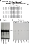
Fig. 7. Evidence for a third site in VDR that is required for optimal Hr interaction
A: Schematic diagram of full-length hVDR showing the DNA-binding domain (DBD), the C-terminal extension of the DNA-binding domain (CTE) [Hsieh et al., 1999], and the ligand-binding domain (LBD) of intact VDR. Two Hr interaction domains are located in the ligand binding domain of hVDR between residues 134 and 303. Major Hr site #1 (202–303) was revealed by immunoprecipitation [Hsieh et al., 2003] and minor Hr site #2 (cross-hatched; 134–201) was deduced from GST pull-down results (data not shown). The lower portion of panel A depicts the location of conserved clusters of charged residues, which were mutated previously [Hsieh et al., 2003] and in the present study in an attempt to determine if a third Hr binding site exists in VDR, one more proximal to the DNA binding domain. In the first mutant, glutamate residues at positions 98 and 99 in human VDR were changed to lysines to produce E98K/E99K. Next, clusters of basic residues at positions 102–104 and at 109–111 were altered to alanines to produce RKR->AAA 102–104 and KRK->AAA 109–111, respectively. The lower portion of panel A illustrates conservation of these residues among VDRs (NR1I1) and also among the related PXR (NR1I2) and CAR (NR1I3) receptors. Sequence sources are as follows: rat VDR NP_058754.1; mouse VDR NP_033530.2; human VDR NP_000367.1; rat PXR NP_443212.1; mouse PXR NP_035066.1; human PXR NP_003880.3; rat CAR NP_075230.1; mouse CAR NP_033933.2; human CAR NP_001070950.1. Human VDR proteins containing mutations in each of the charged clusters were separately expressed in an in vitro transcription-translation system along with wild-type VDR (see Methods). B and C: A representative GST pull-down experiment designed to assess the ability of these mutant proteins to bind immobilized rat Hr. B: Display of 4.5% of the inputs of 35S-labeled wild type (wt) or mutant VDRs used in the pull-down reactions. C: Autoradiograph of a gel containing point-mutated hVDR pull-down reactions using either immobilized GST protein (negative control) or GST-Hr fusion protein containing full-length rat Hr.
Fig. 8
Fig. 8. Integrated model for the functional interactions of VDR and Hr in controlling gene expression to drive the mammalian hair cycle
Functional domains in VDR and Hr are defined in the legends to Fig. 1D, Fig. 2A, and Fig. 7, and in the text. The upper portion depicts schematically the interaction interface between VDR and Hr, which appears to consist of three domains in VDR (denoted in red as Hr sites #1 – #3) and four domains in Hr, previously described [Potter et al., 2001; Moraitis et al., 2002; Potter et al., 2002] as LXXLL-like nuclear receptor interaction domains for either RORα (shaded in green: LCRLL and LCELL) or TR (shaded in blue: LDSII and VSDLI). The central portion illustrates the position in Hr of a set of naturally occurring mutations which confer the alopecic phenotype and were evaluated experimentally in the present study and/or by Wang and coworkers [Wang et al., 2007]. Residue numbers shown in black are for rat Hr; human Hr numbering is shown in magenta underneath the rat numbering. Mutated amino acids in human patients that display alopecia with papular lesions (APL, OMIM #209500) are designated with an asterisk; the corresponding residue in rHr that was evaluated in the current study is shown above the human residue number. A single alopecic Hr mutation from mice, G985W, is represented by the designation G985 with the corresponding human Hr residue number shown in parentheses below. The region of hHr that is encoded by exon 17 and that is missing in a human variant of Hr (isoform b) [Cichon et al., 1998] is shown at upper right with both rat (black) and human (magenta) residue numbering (note that is this isoform is only found in the human). Potential functional interactions between these residues and either HDACs or other unknown nuclear factors are depicted as large arrows in the lower portion and are discussed in the text. Residues 865 to 1204 in rat Hr show high homology to the Jumonji domain-containing protein JmJD1 (34% homology to rat JmJD1B and 33% each to rat JmJD1A and JmJD1C; for accession numbers used in these comparisons see legend to Fig. 3); this region of rat Hr is shaded yellow. Most of the alopecia conferring mutants occur in this region of Hr, as discussed in the text, and lead us to propose that the C-terminal JmJD1-homologous domain in Hr is enzymatically active in histone demethylation, yielding chromatin modifications that repress the expression of genes to control the mammalian hair cycle. The proposed histone demethylase activity of Hr likely operates in concert with histone deacetylases recruited by the JmJD1-homologous domain as well as other repressive domains of Hr to complete a constellation of chromatin repressive alterations required to silence RXR-VDR-Hr target genes. Potential interactions between these residues or regions and either HDACs or other unknown nuclear factors are discussed in the text.

Similar articles

Cited by

References

    1. Ahmad W, Faiyaz ul Haque M, Brancolini V, Tsou HC, ul Haque S, Lam H, Aita VM, Owen J, deBlaquiere M, Frank J, Cserhalmi-Friedman PB, Leask A, McGrath JA, Peacocke M, Ahmad M, Ott J, Christiano AM. Alopecia universalis associated with a mutation in the human hairless gene. Science. 1998;279:720–724. - PubMed
    1. Ahmad W, Zlotogorski A, Panteleyev AA, Lam H, Ahmad M, ul Haque MF, Abdallah HM, Dragan L, Christiano AM. Genomic organization of the human hairless gene (HR) and identification of a mutation underlying congenital atrichia in an Arab Palestinian family. Genomics. 1999;56:141–148. - PubMed
    1. Aita VM, Ahmad W, Panteleyev AA, Kozlowska U, Kozlowska A, Gilliam TC, Jablonska S, Christiano AM. A novel missense mutation (C622G) in the zinc-finger domain of the human hairless gene associated with congenital atrichia with papular lesions. Exp Dermatol. 2000;9:157–162. - PubMed
    1. Bergman R, Schein-Goldshmid R, Hochberg Z, Ben-Izhak O, Sprecher E. The alopecias associated with vitamin D-dependent rickets type IIA and with hairless gene mutations: a comparative clinical, histologic, and immunohistochemical study. Arch Dermatol. 2005;141:343–351. - PubMed
    1. Brancaz MV, Iratni R, Morrison A, Mancini SJ, Marche P, Sundberg J, Nonchev S. A new allele of the mouse hairless gene interferes with Hox/LacZ transgene regulation in hair follicle primordia. Exp Mol Pathol. 2004;76:173–181. - PubMed

Publication types

LinkOut - more resources