

Review
doi: 10.1002/jso.21581.
Sentinel node biopsy in melanoma: technical considerations of the procedure as performed at the John Wayne Cancer Institute
Affiliations
- PMID: 20512942
- PMCID: PMC2879706
- DOI: 10.1002/jso.21581
Item in Clipboard
Review
Sentinel node biopsy in melanoma: technical considerations of the procedure as performed at the John Wayne Cancer Institute
J Surg Oncol.
.
Abstract
Since its first description in 1990, sentinel node (SN) biopsy has become the standard for accurate staging of a melanoma-draining regional lymphatic basin. This minimally invasive, multidisciplinary technique can detect occult metastases by selective sampling and focused pathologic analysis of the first nodes on the afferent lymphatic pathway from a primary cutaneous melanoma. An understanding of preoperative lymphoscintigraphy, intraoperative lymphatic mapping, and the definition of SN are critical for surgical expertise with SN biopsy.
(c) 2010 Wiley-Liss, Inc.
Figures
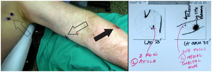
Preoperative lymphoscintigram (right panel) reveals drainage of a forearm melanoma (left panel, solid arrow) to an ectopic cubital node in the upper arm (left panel, open arrow), in addition to expected foci in the axillary basin (left panel, dotted arrow).
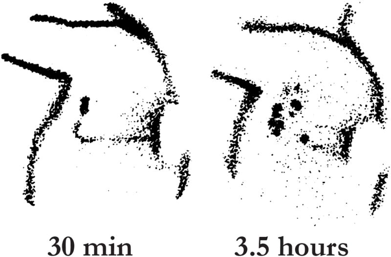
Effect of radiotracer transit time on SN identification. The lymphoscintigram demonstrates the dynamic quality of radiotracers in a patient with melanoma of the left back. The image at 30 minutes depicts the lymphatic channel and SN. Delayed images taken at 3.5 hours show second-tier, non-SNs. Although the SN may not disappear from the lymphoscintigram, its level of radioactivity will decrease with time. Reprinted with permission from Glass EC et al[43].
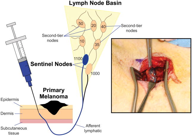
At different times, blue dye and radiotracer are injected intradermally at the site of the primary tumor (radiotracer is injected immediately before preoperative lymphoscintigraphy, and blue dye is injected immediately before the surgical procedure). During intraoperative lymphatic mapping, blue-stained afferent lymphatics are visualized and followed to the first blue-stained node, i.e., the SN (inset). A hand-held gamma probe confirms higher radioactive counts within this SN and can identify additional SNs that are not stained blue.
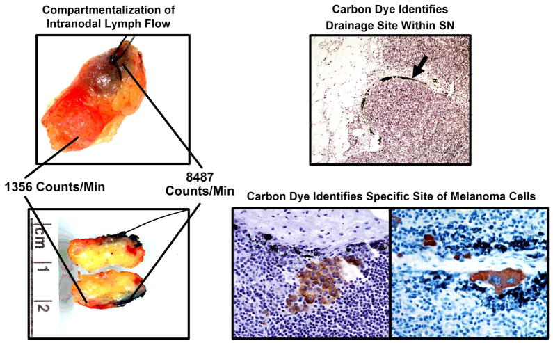
Blue dye and radiotracer identify the first tumor-draining regional lymph nodes (SNs); carbon dye pinpoints the most likely site of tumor cells within these nodes. Unlike blue dye and radioisotopes, carbon is relatively immobile in the node; its intranodal presence therefore can confirm the node as sentinel and help the pathologist identify the most likely intranodal site of any micrometastatic foci. Reprinted with permission from Morton et al. [66]
References
-
- Balch CM, Soong SJ, Atkins MB, Buzaid AC, Cascinelli N, Coit DG, Fleming ID, Gershenwald JE, Houghton A, Jr, Kirkwood JM, McMasters KM, Mihm MF, Morton DL, Reintgen DS, Ross MI, Sober A, Thompson JA, Thompson JF. An evidence-based staging system for cutaneous melanoma. CA Cancer J Clin. 2004;54:131–149. - PubMed
-
- Snow H. Melanotic cancerous disease. Lancet. 1892;2:872.
-
- Balch CM, Soong SJ, Murad TM, Ingalls AL, Maddox WA. A multifactorial analysis of melanoma. II. Prognostic factors in patients with stage I (localized) melanoma. Surgery. 1979;86:343–351. - PubMed
-
- Milton GW, Shaw HM, McCarthy WH, Pearson L, Balch CM, Soong SJ. Prophylactic lymph node dissection in clinical stage I cutaneous malignant melanoma: results of surgical treatment in 1319 patients. Br J Surg. 1982;69:108–111. - PubMed
Publication types
MeSH terms
Grants and funding
LinkOut - more resources
Full Text Sources
Medical
