. 2010 Jun;19(6):1191-204.
doi: 10.1002/pro.396.

The use of native cation-exchange chromatography to study aggregation and phase separation of monoclonal antibodies

Affiliations

The use of native cation-exchange chromatography to study aggregation and phase separation of monoclonal antibodies

Shuang Chen et al. Protein Sci. 2010 Jun.

Abstract

This study introduces a novel analytical approach for studying aggregation and phase separation of monoclonal antibodies (mAbs). The approach is based on using analytical scale cation-exchange chromatography (CEX) for measuring the loss of soluble monomer in the case of individual and mixed protein solutions. Native CEX outperforms traditional size-exclusion chromatography in separating complex protein mixtures, offering an easy way to assess mAb aggregation propensity. Different IgG1 and IgG2 molecules were tested individually and in mixtures consisting of up to four protein molecules. Antibody aggregation was induced by four different stress factors: high temperature, low pH, addition of fatty acids, and rigorous agitation. The extent of aggregation was determined from the amount of monomeric protein remaining in solution after stress. Consequently, it was possible to address the role of specific mAb regions in antibody aggregation by co-incubating Fab and Fc fragments with their respective full-length molecules. Our results revealed that the relative contribution of Fab and Fc regions in mAb aggregation is strongly dependent on pH and the stress factor applied. In addition, the CEX-based approach was used to study reversible protein precipitation due to phase separation, which demonstrated its use for a broader range of protein-protein association phenomena. In all cases, the role of Fab and Fc was clearly dissected, providing important information for engineering more stable mAb-based therapeutics.


Figures
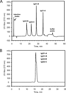
Figure 1
Figure 1
Comparative analysis of a four-mAb mixture by (A) cation-exchange chromatography at pH 5.2 and (B) size-exclusion chromatography at pH 6.8. The antibody mixture contained IgG1-A, IgG1-B, IgG2-B, and IgG2-C.
Figure 2
Figure 2
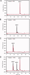
Temperature-induced mAb aggregation in 20 mM sodium phosphate (pH 6.8). Individual and mixed mAb solutions were incubated at 70°C for 10 min without agitation. Control (25°C) and stressed (70°C) samples are shown in black and red, respectively. CEX52 data for individual mAbs are shown for IgG1-A (A), IgG2-A (B), and IgG2-B (C). CEX52 data for a mixture containing IgG1-A, IgG2-A, and IgG2-B are shown in (D).
Figure 3
Figure 3
DSC thermograms in 20 mM sodium phosphate (pH 6.8). A: DSC results for IgG1-A (solid line), IgG2-A (dashed line), and IgG2-B (dashed and dotted line). B: DSC results for intact IgG1-B (solid line) and its Fab (dashed line) and Fc (dashed and dotted line) fragments. The thermograms correspond to equimolar protein concentrations.
Figure 4
Figure 4
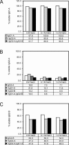
Relative percentage (%) of soluble (A) IgG1-A, (B) IgG2-A, and (C) IgG2-B remaining in the supernatant after a 70°C heat treatment. The bar graphs correspond to temperature-induced aggregation experiments in 20 mM sodium phosphate (pH 6.8) supplemented with 0M, 0.1M, or 1.0M NaCl. The various mAb combinations are shaded differently; the experimental errors have not been determined.
Figure 5
Figure 5
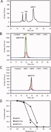
Identification of aggregation-prone regions in temperature-induced mAb aggregation. A: CEX52 separation of papain-digested IgG2-A, illustrating the incomplete digestion of the molecule into constituent Fab and Fc fragments. B: Temperature dependence of IgG2-A Fab precipitation. C: Temperature dependence of IgG2-A Fc precipitation. D: Temperature-dependent aggregation of intact IgG2-A and its Fab and Fc fragments.
Figure 6
Figure 6
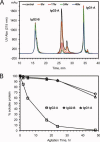
Agitation-induced mAb aggregation in 20 mM sodium phosphate (pH 6.8). A mixture of IgG1-A, IgG2-A, and IgG2-B was rigorously agitated at ambient temperature for up to 48 h. A: CEX52 chromatograms corresponding to different time points during agitation. The species eluting at ∼ 28 min is contained in the IgG2-A drug substance material. The minor peaks eluting at ∼ 36 min are components of the IgG1-A drug substance material. B: Real-time agitation-induced aggregation of the IgG1-A (triangles), IgG2-A (squares), and IgG2-B (circles) mixture.
Figure 7
Figure 7
Acid-induced mAb aggregation in 100 mM sodium acetate (pH 3.5). Individual and mixed mAb solutions were incubated at 4°C and 25°C for up to 48 h without agitation. Control samples (black) were prepared in 20 mM sodium phosphate (pH 6.8) and incubated at 4°C. Acid-treated samples are shown in blue and red for 4°C and 25°C, respectively. CEX52 results after a 48-h incubation of individual IgG1-A (A), IgG2-A (B), and IgG2-B (C) are shown. Corresponding data for a mixture containing IgG1-A, IgG2-A, and IgG2-B are shown in (D).
Figure 8
Figure 8
Fatty acid-induced mAb aggregation. A mixture of IgG1-A, IgG2-A, and IgG2-A Fab and Fc fragments was subjected to precipitation with caprylic acid (red) and heptanoic acid (black) and analyzed by CEX52. The control without fatty acids is shown in blue. The species eluting at ∼ 28 min is contained in the IgG2-A drug substance material. The minor peaks eluting at ∼ 36 min are components of the IgG1-A drug substance material. Note the close correspondence between the loss of intact IgG2-A and its Fc fragment.
Figure 9
Figure 9
Phase separation of mAbs. A: CEX52 analysis of acid-treated and neutralized Protein A-purified IgG2-A from the precipitate (solid line) and supernatant (dashed line). As protein concentration in the precipitate was ∼ 6 times higher than in the supernatant (94.1 ± 1.6 mg/mL vs. 16.2 ± 0.8 mg/mL), the data for the supernatant was normalized with respect to the pellet. Note the differences in homogeneity of precipitated IgG2-A compared to its soluble fraction. B: Protein partitioning experiment on acid-treated and neutralized Protein A-purified IgG2-A. Native IgG2-A Fab and Fc fragments were added to IgG2-A at low quantities before sample refrigeration (see text). Illustrated are four CEX52 traces corresponding to supernatant (blue) and pellet (green) formed in the presence of Fab, and supernatant (black) and pellet (red) formed in the presence of Fc. Note the equal recovery of the Fc fragment from the supernatant and the pellet (the black trace is obscured by the red trace), indicating a lack of preferential partitioning into the solid phase.

References

    1. Cleland JL, Powell MF, Shire SJ. The development of stable protein formulations: a close look at protein aggregation, deamidation, and oxidation. Crit Rev Ther Drug Carrier Syst. 1993;10:307–377. - PubMed
    1. Wang W. Protein aggregation and its inhibition in biopharmaceutics. Int J Pharm. 2005;289:1–30. - PubMed
    1. Cromwell ME, Hilario E, Jacobson F. Protein aggregation and bioprocessing. AAPS J. 2006;8:E572–E579. - PMC - PubMed
    1. Weiss WF, IV, Young TM, Roberts CJ. Principles, approaches, and challenges for predicting protein aggregation rates and shelf life. J Pharm Sci. 2009;98:1246–1277. - PubMed
    1. Roberts CJ. Non-native protein aggregation: pathways, kinetics, and shelf-life prediction. In: Murphy RM, Tsai A, editors. Misbehaving proteins: protein (mis)folding, aggregation, and stability. New York: Springer; 2006. pp. 17–46.

MeSH terms

LinkOut - more resources